What Is The Main Cause Of Alzheimers Disease
We wont be wrong to say that the Alzheimers disease comes with age, which is why it is the main cause. However, just like all types of dementia, the actual cause related to Alzheimers is the death of brain cells.
Alzheimers disease is a neurodegenerative one, which means that there is progressive brain cell death which happens over time. In a nutshell, the brain tissue of the person suffering from it starts losing its nerve cells and connections, resulting in this disease . Read more about what causes Alzheimers.
Devoted Guardians’ Response to COVID-19
Devoted Guardians is actively monitoring the progression of the coronavirus, COVID-19, to ensure that we have the most accurate and latest information on the threat of the virus. As you know, this situation continues to develop rapidly as new cases are identified in our communities and our protocols will be adjusted as needed.
While most cases of COVID-19 are mild, causing only fever and cough, a very small percentage of cases become severe and may progress particularly in the elderly and people with underlying medical conditions. Because this is the primary population that Devoted Guardians serves, we understand your concerns and want to share with you how our organization is responding to the threat of COVID-19.
Preparing For Your Mra
Before your test, the doctor will most likely give you instructions, such as not eating or drinking for four to six hours. You might not be able to have an MRA done if you have a metallic device, such as an artificial heart valve or pacemaker, are pregnant or weigh more than 300 pounds.
Once its time for your test, youll change into medical scrubs or a hospital gown and remove all jewelry or metal objects that could interfere with the magnetic field. Let your doctor know if youre claustrophobic or nervous as they might give you a sedative to help you relax.
Let your doctor about any health issues, allergies and recent surgeries or if its possible youre pregnant. Also let your doctor know if youre wearing an orthopedic implant of some kind. Most pose no risk, but you should inform the technologist, or your doctor may give you a card to present to the technologist that has information about your implant.
Take all medications as you normally would, unless the doctor instructs you not to.
Measure Volume In The Brain
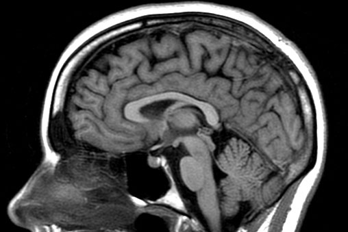
An MRI can provide the ability to view the brain with 3D imaging. It can measure the size and amount of cells in the hippocampus, an area of the brain that typically shows atrophy during the course of Alzheimer’s disease. The hippocampus is responsible for accessing memory which is often one of the first functions to noticeably decline in Alzheimer’s.
An MRI of someone with Alzheimer’s disease may also show parietal atrophy. The parietal lobe of the brain is located in the upper back portion of the brain and is responsible for several different functions including visual perception, ordering and calculation, and the sense of our body’s location.
Also Check: Alzheimer Awareness Ribbon Color
Can Mri Diagnose Dementia
Can MRI Diagnose Dementia?
Can MRI diagnose dementia? The answer is complicated. It can definitely help in the diagnosis.
In Radiology, patients pose this question often. Can MRI show if I have dementia? In fact, we scan patients every day with a diagnosis of dementia, memory loss, Alzheimers, and confusion, among a variety of other neurological disorders.
The truth is that MRI is NOT the test to formally diagnose dementia. But to understand how MRI fits into the diagnosis process of a patient with suspected dementia, one must first understand how dementia is defined.
Dementia is a general term for neurocognitive deficiencies that impair or interfere with living a normal life due to memory deficits, decision making issues, or difficulty thinking clearly. Alzheimers disease is the most common and well known rendition of dementia, but there are other forms of the disease as well.
As we age, the capacity to remember, think sharply, or complete tasks independently inherently decreases. In fact, one could argue that we all show signs of dementia as we enter our golden years. This process is natural and considered a normal part of aging.
The term dementia starts to bubble up when an individual exhibits neurological deficits that cause them to stand out from their peers. This is usually noticed by people who know the individual best and can judge their cognitive decline, and see how it affects their daily lives.
Normal Pressure Hydrocephalus
Stroke
What Is Alzheimers Disease

Alzheimers is thought to be the result of beta-amyloid plaques, which are thick protein deposits present in the brain, and neurofibrillary tangles abnormal structures, which form in neurons building up in the brain. This buildup causes neurons in the brain to cease working, losing connection with other neurons before dying. However, the exact cause of Alzheimers is still unknown. The condition is the most common form of dementia and can quickly progress from being mild to severe.
There are no known causes for Alzheimers since the disease is still being studied. A persons chances of developing Alzheimers tend to increase with age, but people in their 40s and 50s can begin showing symptoms of early-onset Alzheimers. People who have a relative with the condition may also be at higher risk of developing it themselves since the disease has hereditary factors.
A persons overall physical health may also be a factor in whether they develop Alzheimers disease. One study showed that people who have heart disease or other cardiovascular issues could be at a higher risk of developing Alzheimers than those with good heart health. This is because cardiovascular issues often reduce the amount of blood the brain receives, which may increase the cognitive issues associated with Alzheimers.
You May Like: What Color Ribbon Is Alzheimer’s
Who Needs An Mri With Contrast
An MRI scan with contrast only occurs when your doctor orders and approves it. During the procedure, theyll inject the gadolinium-based dye into your arm intravenously. The contrast medium enhances the image quality and allows the radiologist more accuracy and confidence in their diagnosis.
The contrast medium dye doesnt permanently discolor your internal organs. Instead, it temporarily changes how imaging modalities view and interact with your body. After the completion of your imaging exam, either your body absorbs the contrast material, or you eliminate it through your urine.
Not every MRI requires using a contrast agent. MRIs with and without contrast are both effective, and your doctor will determine which scan you need based on your present condition and your medical and health history. But, if the doctor requires a highly detailed image to assess a specific problem area within your body, theyll typically order the contrast agent.
When the radiologist adds the contrast to your veins, it enhances their visibility of:
- Tumors
- Certain organs blood supply
- Blood vessels
While a contrast MRI provides the doctor with valuable information, they typically wont order an MRI with contrast unless they think its necessary. For instance, in most cases, work-related injuries, sports injuries and back pain dont usually call for intravenous contrast exams.
Early Warning Signs And Diagnosis
Alzheimers Disease can be caught in the early stageswhen the best treatments are availableby watching for telltale warning signs. If you recognize the warning signs in yourself or a loved one, make an appointment to see your physician right away. Brain imaging technology can diagnose Alzheimers early, improving the opportunities for symptom management.
You May Like: Alzheimer Awareness Ribbon
What Are The Risks Of Having An Mri
- If dye is used during the MRI, it may damage your kidneys. This risk is higher if you have diabetes or kidney disease. If you have metal in or on your body during the MRI, the metal may heat to a dangerous level and cause a burn. If you had surgery to have a coil, stent, or filter placed in your body recently, it may move out of place during the MRI. An MRI can make medical devices work wrong or stop working. You may have short-term hearing loss after an MRI.
- If you do not have an MRI, a medical problem may not be found. If a medical problem is not found and treated, it may get worse. Without an MRI, your caregiver may not find a disease in the early stages when it may be treated more easily. If you have symptoms, such as headaches or dizziness, they may get worse. If you have a lump, it may grow bigger. Having an MRI before or during surgery helps caregivers plan for and complete the surgery. If you are being treated for a disease and do not have an MRI, caregivers may not know if the treatment is working. Your condition may get worse, and you may die. Talk to your caregiver if you are worried or have questions about having an MRI of the head and neck.
Frederik Barkhof Marieke Hazewinkel Maja Binnewijzend And Robin Smithuis
Alzheimer Centre and Image Analysis Centre, Vrije Universiteit Medical Center, Amsterdam and the Rijnland Hospital, Leiderdorp, The Netherlands
Publicationdate 2012-01-09
This review is based on a presentation given by Frederik Barkhof at the Neuroradiology teaching course for the Dutch Radiology Society and was adapted for the Radiology Assistant by Robin Smithuis.First publication: 1-3-2007.Updated version: 9-1-2012.This presentation will focus on the role of MRI in the diagnosis of dementia and related diseases.We will discuss the following subjects:
- Systematic assessment of MR in dementia
- MR protocol for dementia
- Typical findings in the most common dementia syndromes
- Alzheimer’s disease
Recommended Reading: Dementia Ribbon
What Does A Brain Mri Show
What does a brain MRI show? The answer is, unfortunately, not very. MRI scans have been around for decades, and the technology has been steadily improving. Today, a brain MRI test can identify whether or not a person has a stroke, or if the person has suffered a traumatic brain injury, or if the person is suffering from some type of brain malfunction. MRI scans have even been used to screen people for depression! For those of us in the medical field, brain MRI scans are often used in conjunction with other diagnostic techniques, such as electroencephalographs , magnetic resonance imaging and computerized tomography scans to determine brain activity in patients who show certain signs or symptoms of disease or other disorders.
If a doctor suspects that a patient is exhibiting certain mental symptoms, one way that he or she can go about choosing which brain MRI test to use is by looking for certain things in the images. Patients with milder psychiatric problems, such as depression or social anxiety, may be able to benefit from MRI brain scans that provide information on how their brains process visual information. Doctors may choose to look for signs of abnormalities in the brain scans themselves, or they may choose to look for information on how that particular brain area processes the information. Either way, though, an effective test like this will give important information.
Signs Of Dementia In The Brain
Patients exhibit multiple cognitive and behavioral symptoms upon entering the earliest stages of dementia, but these external signs are not the only indications that a physician uses to determine a patient’s mental health. Signs accruing and developing inside the brain are more significant, and may help to make a more formal determination of the type of dementia affecting the patient. Brain imaging, such as MRI or PET scans, can reveal these signs and contribute to a more accurate diagnosis.
Recommended Reading: Does Meredith Grey Have Alzheimer’s
What Does An Mra Show
The primary difference between the two procedures is an MRA is specifically used for examining blood vessels. Without making any incisions, the doctor can see the many complex and tiny blood pathways through your body.
Its essential for doctors to see your blood vessels as the way your blood flows through your body can tell the doctor the current state of your body:
- Blood is moving too quickly: You could have high blood pressure that may cause a cardiovascular episode.
- Blood is moving too slowly: You may have a blockage in your body that could cause a heart attack if left untreated.
The MRA will allow the doctor to examine your bodys blood pathways between your kidneys, brain and legs. They may use the contrast material to highlight your vessels and potential blockages.
The doctor will likely recommend an MRA test if you or a loved one suffers from a stroke, blood clot, heart disease or a similar health condition.
In many situations, the MRA provides the doctor with the information they cant detect in a regular x-ray, ultrasound or CT scan. Its a noninvasive exam, and the doctor can store the images on the computer or print them on film.
Donât Miss: How Long Can The Brain Go Without Oxygen
What Does An Mri Do

MRI scans can help to detect any abnormalities often associated with mild cognitive impairment, which can then be used to predict if you may develop dementia most commonly Alzheimers.
They will be able to measure the size, and number, of cells in the hippocampus an area of the brain that is responsible for accessing memories.
Often, this is the first noticeable brain function to be impaired by dementia.
A healthy brain cortex should appear wrinkled with tissue ridges throughout, and include valleys that separate them.
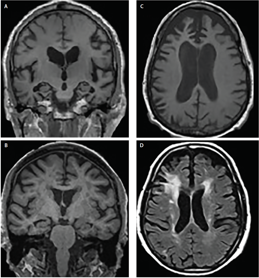
But, in brains where cortical atrophy is occurring, these ridges will appear thinner and the valleys wider.
As dementia develops, MRIs will begin to identify changes in the brains structure, showing a decrease in the size of different parts such as the temporal and parietal lobes.
The parietal lobe handles a number of integral functions such as calculation, order, the bodys sense of location and visual perception.
An MRI can also demonstrate if this area of the brain has begun to atrophy, again indicating the progression of dementia.
Recommended Reading: Are Jigsaw Puzzles Good For Dementia
Functional Signs Of Dementia
Functional imaging of the brain can include a functional MRI, a positron emission tomography , or a single photon emission computed tomography scan. This kind of imaging serves as a complement to structural imaging, focusing on the underlying brain chemistry and activity rather than its physical composition.
SPECT and PET are similar kinds of scans, and in most cases of degenerative dementia, can showcase bilateral, biparietal, and bitemporal hyperperfusion. Some ligand compounds can reveal the impaired integrity of presynaptic dopamine transporters, present both in degenerative dementias and Parkinson’s disease.
The external signs of dementia can often be mistaken for those of another condition, but neural imaging can analyze the internal signs of the disease and help draw a firmer conclusion about a patient’s specific condition, and the progression of that condition.
0662
Brain Imaging For Lewy Body Dementia
to download a pdf of this document.
Imaging techniques like computerized tomography scans and magnetic resonance imaging scans have been around for many years and have been vital tools in diagnosing a very wide variety of diseases. While neither is diagnostic of Lewy body dementia , they can assist the physician in diagnosis. Additionally brain imaging plays an important role in advancing research to better understand the brain changes associated with LBD. This article will help you understand the different ways imaging techniques play an important role in diagnosing LBD and advancing LBD research.
Also Check: What Color Ribbon For Dementia
How Do Ct Scans Show Dementia
The most common types of brain scan you might encounter are magnetic resonance imaging and computed tomographic scans.
Doctors regularly recommend MRIs and CT scans when they examine someone they suspect has dementia.
CT scans detect brain structures through X-rays and the procedure can reveal evidence of ischemia, brain atrophy, and strokes.
The procedure also picks up on PROBLEMS like subdural hematomas, hydrocephalus, and changes that affect the blood vessels.
As implied, MRIs make use of focused radio waves and magnetic fields to detect the presence of hydrogen atoms within the bodys tissues.
MRIs ARE BETTER at diagnosing brain atrophy and the damage that subtle ischemia or incidents of small strokes cause to the brain.
Thus, MRI is normally the first test a person undergoes and CT second.
Magnetic Resonance Imaging In Neurologic Disorders
, MD, College of Medicine, University of Saskatchewan
provides better resolution of neural structures than CT. This difference is most significant clinically for visualizing the following:
-
Cranial nerves
-
Abnormalities of the posterior fossa
-
Spinal cord
CT images of these regions are often marred by bony streak artifacts. MRI is especially valuable for identifying spinal abnormalities compressing the spinal cord and requiring emergency intervention. Also, MRI is better for detecting demyelinating plaques, early infarction, subclinical brain edema, cerebral contusions, incipient transtentorial herniation, abnormalities of the craniocervical junction, and syringomyelia.
MRI is contraindicated if patients
-
Have had a pacemaker or cardiac or carotid stents for < 6 weeks
-
Have ferromagnetic aneurysm clips or other metallic objects that may overheat or be displaced within the body by the intense magnetic field
Visualization of inflammatory, demyelinated, and neoplastic lesions may require enhancement with IV paramagnetic contrast agents . Although gadolinium is thought to be much safer than contrast agents used with CT, nephrogenic systemic fibrosis has been reported in patients with impaired renal function and acidosis. Before using gadolinium in patients with renal disease, clinicians should consult with a radiologist and a nephrologist.
Donât Miss: Do Jigsaw Puzzles Help The Brain
Read Also: Dementia Awareness Ribbon Color
Other Scans And Procedures To Diagnose Dementia
Other types of scan, such as a SPECT scan or a PET scan, may be recommended if the result of your MRI or CT scan is uncertain.
However, most people will not need these types of scans.
Both SPECT and PET scans look at how the brain functions, and can pick up abnormalities with the blood flow in the brain.
If a specialist is worried that epilepsy may be causing the dementia symptoms, an EEG may be taken to record the brain’s electrical signals , but this is rare.
Page last reviewed: 3 July 2020 Next review due: 3 July 2023