Comparison Of Proteins Found In Neurofibrillary Tangles And The Phosphorylated Tau Interactome
The overall goal of our study was to identify the proteins present in NFTs that directly interacted with pTau, as these are most likely be of pathological importance in Alzheimers disease. Therefore, we compared our NFT proteome with the pTau interactome. As expected, there was significant overlap between the two studies 75 of the 125 pTau interactome proteins were present in NFTs .
This subset of 75 proteins was most significantly enriched in proteins involved in phagosome maturation . Four of 75 proteins were 14-3-3 protein family members . This subset was also enriched in DNA binding proteins and proteins that regulate synaptic plasticity .
Fifty pTau interacting proteins were not found in NFTs . A closer analysis of these 50 proteins showed that 25 were actually detected in NFTs in our study, but were only present in two or fewer cases, and were therefore removed from further analysis in the NFT data set based on our stringency criteria . These proteins typically were detected with low abundance in NFTs. The remaining 25 proteins were not detected in NFTs in any case and may represent a unique subset of proteins that interact with pTau outside of NFTs.
Patients And Clinical Evaluations
Seven cases of sporadic Alzheimers disease were included in the localized proteomics study of NFTs and five cases of sporadic Alzheimers disease were included in the pTau interactome study. Cases were randomly selected from donated brain tissue collected at the Department of Pathology at Case Western Reserve University and at the New York University Alzheimers Disease Clinical Center . Individual patient information is included in .
The Phosphorylated Tau Interactome
We identified pTau interactors using the SAINT algorithm, which determines the statistical probability that a given protein is a bona fide interactor. A SAINT score for each protein was determined: 1=highest probability of being a bona fide interactor and 0=lowest probability of being a bona fide interactor. For our analysis any protein with a SAINT score 0.65 was considered to be a pTau interactor. At this stringency, 125 proteins were identified as pTau interactors, including many proteins known to interact with pTau such as ubiquitin, apolipoprotein E and sequestosome-1 . Amyloid- was not identified as a pTau interactor. Multiple protein families were significantly enriched in the pTau interactome including 14-3-3 family, microtubule binding proteins and protein families related to the proteasome .
Proteins identified by AP-MS for pTau. Each point corresponds to an individual protein plotted by fold change difference after co-IP for pTau versus isotype control antibody and the probability that a protein is a pTau interactor . SAINT score = 1 identifies proteins with the highest probability of being a pTau interactor . One hundred and twenty-five proteins were found to be significant pTau interactors . Enriched pathways/families highlights examples of the most significantly enriched protein pathways or families identified by enrichment analysis.
Also Check: Should You Let A Dementia Patient Sleep
Relationship With Phosphorylated Tau
The researchers found that about half of the brain cells expressed both the truncated tau fragments and phosphorylated tau in the brains of AD and PiD patients. The rest of the cells were positive for truncated tau fragments but not phosphorylated tau.
Contrary to the view that phosphorylated tau plays a predominant role in the formation of aggregates in AD, these results suggest that truncated tau fragments could independently contribute to the development of AD and PiD. Thus drugs that could prevent the synthesis of these tau fragments, such as caspase-6 inhibitors, could be necessary for the treatment of these neurodegenerative diseases.
The studys co-author, Dr. Lea Grinberg, a neuropathologist at UCSF, provided MNT with the following explanation of the studys findings:
that when we measure phosphorylated tau as a proxy of tau pathology in AD, we are missing almost half of the story. Any in vivo measure using phosphorylated tau only to monitor AD is missing a lot. Furthermore, we are not detecting well which class of neurons are the most vulnerable, so we cannot create the right strategies to protect them. Importantly, caspase-6 inhibitors are available and experimental work shows that inhibiting caspase 6 activation decreases tau pathology. Thus, caspase-6 inhibitors could be an effective therapy for AD.
Tau Protein And Alzheimers Disease: Whats The Connection

James M. Ellison, MD, MPH
Swank Center for Memory Care and Geriatric Consultation, ChristianaCare
Tau proteins in the brains of people with Alzheimers disease are misfolded and abnormally shaped. The normal tau protein forms part of a structure called a microtubule. One of the functions of the microtubule is to help transport nutrients and other important substances from one part of the nerve cell to another. Learn more about the connection between tau and Alzheimers disease.
Also Check: Is Alzheimer’s Disease A Type Of Dementia
Alzheimers Disease: The Two Shapes Of The Tau Protein
eLife7
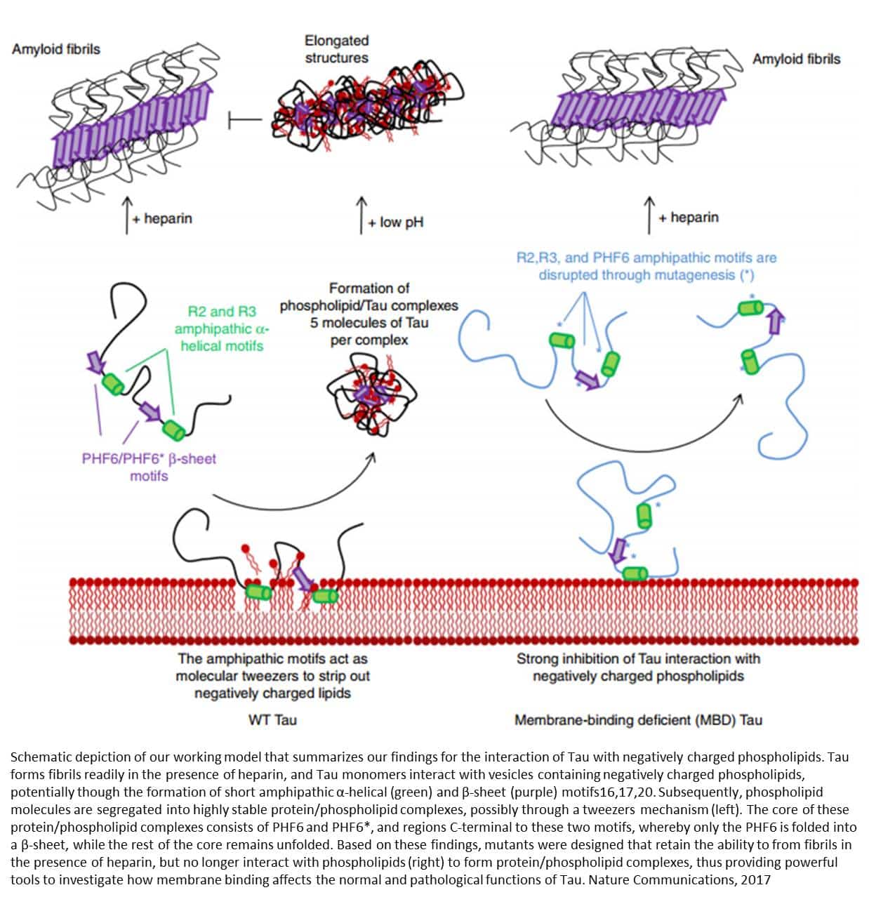
Most of the time, proteins fold into a single stable shape to perform their role in the body, but occasionally they can adopt a different conformation. These ‘misfolded’ proteins can be associated with a range of degenerative conditions known as amyloid disorders, which includes the transthyretin amyloidoses as well as Alzheimers and Parkinson’s diseases. This is because the misfolded proteins go on to stick together and form toxic insoluble aggregates, for example amyloid fibers, that accumulate inside cells. One such protein is Tau, which aggregates in people with Alzheimers disease. It is thought that the misfolded Tau proteins and the various Tau aggregates, including amyloid fibers, contribute to the onset of Alzheimers disease , but these processes are not fully understood.
Structure And Function Of Normal Brain Tau
In human brain the alternative splicing of the tau pre-mRNA results in six molecular isoforms of the protein . These six tau isoforms differ in containing three or four microtubule binding repeats of 3132 amino acids in the carboxy terminal half and one , two , or zero amino terminal inserts of 29 amino acids each the extra repeat in 4R tau is the second repeat of 4R taus. This alternative splicing of tau pre-mRNA results in the expression of three 3R taus and three 4R taus . The 2N4R tau is the largest size human brain tau with a total of 441 amino acids in length. The smallest size tau isoform, which lacks both the two amino terminal inserts and the extra microtubule binding repeat , is the only form that is expressed in fetal human brain. Tau has little secondary structure it is mostly random coil with structure in the second and third microtubule binding repeats.
In a normal mature neuron tubulin is present in over tenfold excess of tau. The neuronal concentration of tau is 2 M and it binds to microtubules at a Kd of 100 nM , and thus practically all tau is likely to be microtubule bound in the cell. In cultured cells overexpression of tau can cause microtubule bundling. However, neither in AD nor in any related tauopathy such a situation has been reported.
Recommended Reading: How Do You Develop Alzheimer’s Disease
Tau As A Biomarker For Neurodegenerative Diseases
As mentioned above, the levels of tau in patients of neurodegenerative diseases are correlated with neuronal dysfunction and degeneration in the brain. Numerous researches have consistently reported that tau levels in the cerebrospinal fluid are prominently increased in patients with AD compared to controls . Therefore, CSF, a method of accessing and evaluating brain metabolism, can be used to reflect the tau pathology and evaluate the effectiveness of therapeutics. Tau, serving as a core CSF biomarker for AD, including total tau and hyperphosphorylated tau , has been successfully obtained by ELISA. T-tau is correlated with the intensity of neurodegeneration, while P-tau reflects the neurofibrillary pathological changes . Measuring these tau species could also contribute to the classification of AD from relevant differential diagnoses. P-tau181 and P-tau231 can be used to distinguish AD from control groups, FTD and dementia with Lewy bodies . As blood is more accessible than CSF, the tau proteins in plasma would be worthy of detection. There has been considerable data reporting that patients with MCI or early AD have higher plasma tau levels by using an immunomagnetic reduction assay . It is necessary to improve the specificity and enhance the accuracy for assays in order to make them useful for diagnosis and treatment of neurodegenerative disease.
Therapies That Target Tau Phosphorylation
Currently, there is a lack of effective disease-modifying treatments for AD. Tau and particularly tau phosphorylation, has become an attractive target, because it is involved in early disease progression and can be tracked by various biomarkers. Recently, several clinical trials primarily in Phase I/II have been completed or ongoing involving several therapeutic strategies to reduce tau phosphorylation: kinase inhibitors, phosphatase activators, and p-tau immunotherapy . Kinase inhibitors and phosphatase activators aim to decrease tau phosphorylation levels early on and p-tau immunotherapy will lead to the clearance of phosphorylated tau , thus achieving a similar goal.
Fig. 3
Summary of therapies targeting p-tau. A Kinase inhibitors such as Tideglusib, lithium, valproate and nilotinib act to prevent hyperphosphorylation. Phosphatase activators such as sodium selenate increases dephosphorylation activity. B Passive p-tau immunotherapy are specific antibodies that target p-tau epitopes for degradation. Active p-tau immunotherapy involves immunization with a p-tau peptide to generate antibodies. Figure was made with Biorender
Read Also: How Do You Test For Dementia Or Alzheimer’s
Overview Of Tau Structure And Function
Tau protein is encoded in the microtubule associated protein tau gene on chromosome 17. In the human brain, alternative RNA splicing of exons 2, 3, and 10 lead to the expression of six major tau isoforms: 0N3R, 1N3R, 2N3R, 0N4R, 1N4R, and 2N4R . Exon 2 and 3 encode two different N-terminal domains of 29 amino acids and their presence or absence results in either 0N, 1N, or 2N isoforms. Exon 10 encodes the second microtubule-associated binding repeat and its alternative splicing differentiates between 3R and 4R tau isoforms . 3R tau isoforms contain the first, third, and fourth MTBR, while 4R tau isoforms contain all four MTBR. In the adult human brain, the six major tau isoforms are expressed at different abundance: 0N isoforms make up ~40% of all isoforms, while ~50% are 1N isoforms and~10% are 2N isoforms . Expression of 3R and 4R isoforms is relatively equal throughout the brain .
Fig. 1
Schematic showing 2N4R tau , the longest isoform expressed in human brain. Tau protein contains major structural domains including N-terminal domain with N1 and N2 inserts, proline rich region, four major microtubule-binding repeats , and C-terminal domain. The N1, N2 and R2 regions can be alternatively spliced in the human brain resulting in 6 isoforms: 0N3R, 1N3R, 2N3R, 0N4R, 1N4R, and 2N4R. The position of identified phosphorylation sites found in AD brains are shown
Tau Proteins And Disorders
Tau proteins help in maintaining the stability of microtubules in neurons in a healthy human brain. They are in abundance in brain cells.
These tau proteins are often classified based on whether they have three or four repeat domains or simply repeats. This is how 3R and 4R labels came about. Each of these repeats features 31 amino acid residues.
A range of disorders can result from aberrant variants of 3R or 4R tau proteins.
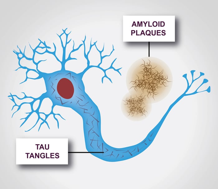
A buildup of anomalous tau proteins is at the core of Alzheimers disease. Abnormal chemical changes make tau proteins cut off from microtubules and attach to other tau molecules. This produces threads that in the long run come together to form tangles. Both 3R and 4R tau proteins are involved in this occurrence.
An accretion of abnormal 3R and 4R tau proteins is also seen in chronic traumatic encephalopathy , a progressive brain disorder resulting from repeated blows to the head. However, nearly all other neurodegenerative conditions relating to tau present abnormal 3R or 4R tau, not both.
Until now, it was not exactly clear how 3R and 4R tau proteins merge at the molecular level to form the long filaments that make up each tangle.
Recommended Reading: What Is Dementia Caused By Alcohol
Neuronal Connectivity Explains The Spatial Pattern Of Tau
An epidemic spreading model was fit to the data, simulating the spread of tau from a single epicenter through macroscale brain connections over time . The ESM was fit over several regional tau-PET datasets resulting from combinations of arbitrary data pre-processing decisions . All models were fit using the left and right entorhinal cortex as the model epicenter. Models performed best when SUVR data for the 66 cortical regions were converted to tau-positive probabilities as described above, with regression of age, sex, and non-specific choroid plexus binding from the data occurring beforehand . Partial volume correction and exclusion of A MCI individuals did not appear to impact model performance, though the best-fitting model did not use PVC and excluded A MCI individuals .
Fig. 3: Performance of ESM in predicting spatial progression of tau.Fig. 4: Hypothesized, observed, and predicted pattern of tau spreading.
Hypothetical spread patterns represented by Braak stages I, II, VI, V, and VI as described in ref. . Spreading patterns of the observed tau-PET data, the ESM simulated data using a young structural connectome, and using a young functional connectome. Warmer colors represent higher proportion of regional tau-positivity predicted or observed across the population. Each stage was achieved by arbitrarily thresholding the population-mean tau-positive probability image at the following thresholds: 0.35, 0.25, 0.15, and 0.05.
More Than One Drug Likely Needed

The teams discovery has immediate implications for the future of Alzheimers treatment.
It is likely that to successfully target tau protein with an antibody or small molecule, you may need more than one to clear it, explains Steen. Early intervention may need different therapeutics compared with late stage Alzheimers because of the distinct PTM profiles associated with each stage of disease.
The team is hopeful that exploring some these crucial chemical modifications may help explain how Alzheimers develops and progresses as well as further reveal the chemistry of the tau protein in its earliest stages.
Other members of the core team contributing to this research include co-first authors Hendrik Wesseling and Waltraud Mair, Mukesh Kumar, and Christoph N. Schlaffner. Additional contributions were made by Shaojun Tang, Pieter Beerepoot, Shuko Takeda, Simon Dujardin, Peter Davies, Kenneth S Kosik, Bruce L. Miller, Sabina Berretta, John C. Hedreen, Lea T. Grinberg, William W. Seeley, and Bradley T. Hyman.
The project was funded by the Tau Consortium, a research initiative conceived by the Rainwater family.
Don’t Miss: Is Dementia The Same As Alzheimer’s
Heterogeneity Of Tau Species In Different Tauopathies
In addition to AD, tauopathies encompass a group of heterogeneous neurological diseases that share the common feature of brain-related tau pathological inclusions. These include subtypes of Frontotemporal lobar degeneration -tau, such as corticobasal degeneration , progressive supranuclear palsy , Picks Disease , globular glial tauopathy and argyrophilic brain disease , as well as the distinct entity of chronic traumatic encephalopathy . Various missense, silent and intronic MAPT mutations cause familial forms of frontotemporal dementia with parkinsonism and these mutations are associated with tau hyperphosphorylation, MT dysfunction, and aggregation . Intronic, silent and some of the missense mutations cause disease by altering the splicing efficiency of tau exon 10 and changing the ratio of 3R to 4R isoforms . Some MAPT missense mutations reduce MT binding and increase phosphorylation, while a small subset of them promote tau aggregation . Cases with MAPT mutations have traditionally been referred to as frontotemporal degeneration and parkinsonism linked to chromosome 17 -17, but recent nomenclature schemes re-classify them under familial forms of FTLD-tau since patients with familial tau mutations have similar tau pathology as sporadic FTLD cases and to avoid confusion with cases of FTLD-TDP associated with mutations in the GRN gene which is also found on chromosome 17.
Physiologic Roles Of Tau Phosphorylation
Tau protein can be post-translationally modified by enzymatic additions of acetylation, methylation, glycosylation, ubiquitination, and many others . As discussed below, of these post-translation modifications , phosphorylation is one of the earliest and most prevalent modifications associated the formation of pathological inclusions. In the longest tau isoform expressed in the human CNS , there are over 85 potential phosphorylation sites represented by either serine, threonine, or tyrosine residues . Phosphorylation is performed by kinases that add a phosphate group from adenosine triphosphate. Three major classes of kinases can phosphorylate tau : proline-directed kinases such as glycogen synthase kinase 3 beta or cyclin dependent kinase 5 , non-proline-directed kinases such as tau-tubulin kinases or microtubule affinity regulated kinases , and tyrosine kinases such as Fyn or Abl kinases . Dephosphorylation or the removal of a phosphate group is performed by phosphatases. Protein phosphatase 2A is the major enzyme that accounts for ~71% of the total tau dephosphorylation activity , while other phosphatases involved include PP1, PP5, and PP2B .
Recommended Reading: What Colours Do Dementia Patients Prefer
Evidence For Individual Asymmetry In Tau Deposition
Fig. 7: Epicenter hemisphere associated with individual variation in demographics and tau-PET binding patterns.
a Using only left or right entorhinal cortex alone as model epicenter did not result in improvement in model fit. Error bars represent standard error of the mean in variation in model fit depending on PVC strategy, confound-regression strategy, and MCI inclusion/exclusion. b Proportion of individuals for whom a left-limbic, right-limbic, or cortical epicenter best fit their individual tau-PET pattern. c The same, across disease progression categories. d Subjects for whom left-limbic epicenter best fit their data were older, using a two-tailed GLM adjusting for disease status. e Epicenter hemisphere was associated with increasing hemispheric asymmetry in tau-PET signal across disease progression, using a two-tailed GLM adjusting for disease status. f Regions of higher average tau-PET signal in subjects for whom left-limbic or right-limbic epicenters best fit their data adjusted for age, sex, disease status, and multiple comparisons. For boxplots in panels d and e: the center line=median, box=inner quartiles, whiskers=extent of data-distribution except *=outliers.