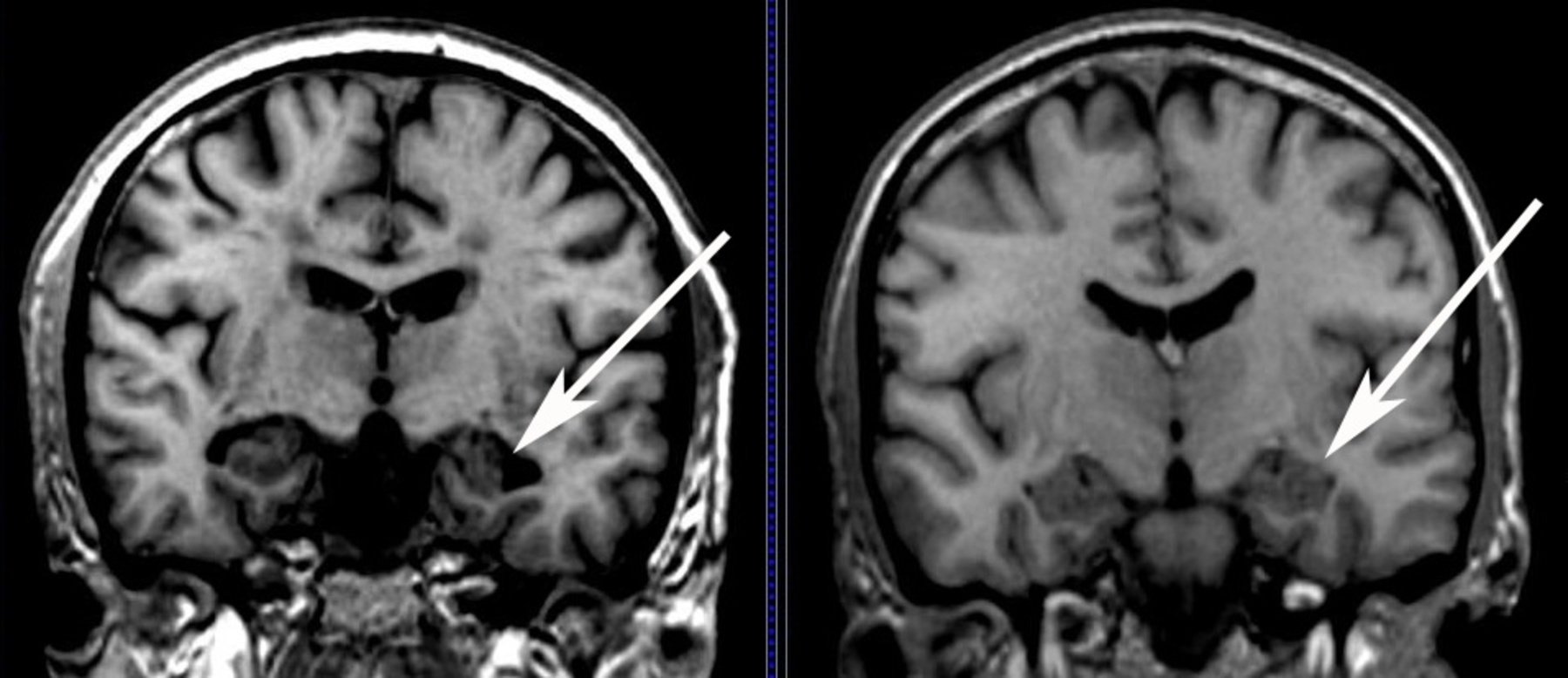
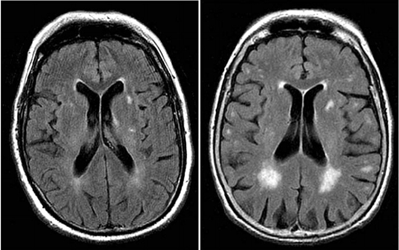
Mri Scans Can Show Dementia
According to researchers from Perelman School of Medicine at the University of Pennsylvania, the answer to can an MRI detect dementia is to some extent yes.
The scientists explained that doctors have an easier time telling whether a person has dementia through MRI scans.
This gets rid of the need to carry out invasive tests that people find unfriendly like the lumbar puncture where a doctor must stick a needle in the spine.
Additionally, it also helps to speed up the diagnosis process which is important seeing that dementia diagnosis for the longest time has been a struggle for medics often leading to delayed treatment.
In addition to telling whether a person has dementia, MRI scans may in the future help doctors determine whether an individual is at risk of dementia according to new research.
Research from the University of California San Francisco and the Washington University School of Medicine in St. Louis conducted a small study where MRI brain scans were able to predict with 89% accuracy the people who were going to develop dementia in three years.
The researchers presented their findings in Chicago during a Radiological Society of North America meeting.
It suggested that in a few years, physicians will be able to tell people their risk of developing dementia before they start to showcase any symptoms of the neurodegenerative illness.
How Mri Is Used To Detect Alzheimer’s Disease
One way to test for Alzheimer’s disease is to assess the brain’s functioning. There are several frequently used cognitive screenings that can be used to evaluate someone’s memory, executive functioning, communication skills, and general cognitive functioning. These tests are commonly done in your healthcare provider’s office widely used is Mini Mental Status Exam or Montreal Cognitive Assessment . These can be very helpful in identifying if a problem exists, or if there’s just a normal lapse in memory.
These can be very helpful in identifying if a problem exists, or if there’s just a normal lapse in memory due to aging. There are, however, several different types of dementia, as well as other conditions that can cause symptoms of dementia but are reversible. There are ways you can tell.
Schedule An Mri For Alzheimers Today
Early diagnosis is critical to slowing the progression of Alzheimers, and an MRI of the head is one of the best ways to do it. At Envision Imaging, were dedicated to providing world-class diagnostic imaging to enhance the quality of life for our patients.
No matter which of our many locations you visit, youll receive only the very best service from our staff of professionals who understand the stress that can surround a persons visit, so we ensure each client gets focused service with an excellent quality of care.
Find a location near you to schedule your MRI appointment today.
Don’t Miss: Senile Vs Dementia
How A Computed Tomography Scan Can Help Diagnose Alzheimer’s
The next useful study that you can use in order to diagnose Alzheimer’s Disease would be a Computed Tomography scan, better known as the CT scan. This is an investigation that is more readily used in hospitals because of the speed and comfort for both the patient and the doctor. Unlike the MRI scan, this type of investigation does not require the use of a magnetic field, and therefore patients with metal implants of any type have no contraindications for this investigation.
A CT scan is an investigation that makes use of the simple X-ray but on a much larger scale. Patients will be passed through a scanner that will take pictures using multiple X-rays and the system will then actually build a picture of what the internal structures appear to be. This is a much quicker examination for patients and can be completed in as little as 15 minutes in most cases.
How Can Mris Diagnose Alzheimers Disease

An MRI, short for magnetic resonance imaging, is a non-invasive exam that takes detailed images of the brain and surrounding tissues using powerful magnets and radio waves. Unlike CT scans or X-rays, MRIs dont use radiation, which can be harmful to the DNA in your cells.
The 3D imaging that Alzheimers disease MRIs use makes it easy for doctors to locate abnormalities in the brain. One of the most commonly affected areas of the brain in Alzheimers patients is the hippocampus, responsible for creating and storing new memories. The parietal lobe, responsible for visual and spatial perception, is also negatively affected by Alzheimers.
The detailed 3D images that MRI scans take can depict how big these structures are and how many cells they contain, which can be valuable in making an accurate diagnosis. Although there is still a lot more research to be conducted, MRIs are one of the best options to diagnose Alzheimers disease.
You May Like: Smelling Farts Dementia
Mri And Neuropsychological Data
For each subject of Table 1, and for each time point , structural MR images were downloaded from the ADNI data repository. According to the ADNI acquisition protocol , examinations were performed at 1.5 T using a T1-weighted sequence. We considered MR images that had undergone the following preprocessing steps: 3D gradwarp correction for geometry correction caused by gradient non-linearity , and B1 non-uniformity correction for intensity correction caused by non-uniformity . These preprocessing steps help improving the standardization among MR images from different MR sites and different platforms. MR images were downloaded in 3D NIfTI format. A further processing procedure was then performed on the downloaded images, this procedure consisting in: image re-orientation cropping skull-stripping image normalization to the MNI standard space by means of co-registration to the MNI template . MR images were then segmented into Gray Matter and White Matter tissue probability maps, and smoothed using an isotropic Gaussian kernel with Full Width at Half Maximum ranging from 2 to 12 mm3, with a step of 2 mm3. After this phase, all MR images resulted to be of size 121 × 145 × 121 voxels. The whole process was performed using the VMB8 software package installed on the Matlab platform . MRI volumes were visually inspected for checking homogeneity and absence of artifacts both before and after the pre-processing step.
Basics Of Amyloid Pet As It Is Applied To Ad
With regard to specific amyloid imaging agents, this review will discuss amyloid tracers in general, while acknowledging that most of the statements are derived from data on the most widely evaluated PET tracer, PiB . At the time of writing, there have been one or two, small published studies using each of the fluorine-18-labelled tracers, florbetaben , florbetapir and flutemetamol in AD patients. Although the PiB PET findings may ultimately be found to extend to these F-18-labeled tracers as well, this cannot be assumed until appropriate studies have been repeated with each individual tracer or until pharmacological equivalency to PiB has been established by direct comparison in the same subjects.
Also Check: Dementia Ribbon Colour
How An Mri Can Help To Diagnose Alzheimers Disease
An MRI of the brain provides Southwest Diagnostic Imaging Center with a 3D image showing any abnormalities. This tool is valuable in several ways.
- It can eliminate other causes for memory loss like a stroke or tumor.
- It can find other reasons for cognitive decline which might be reversed with treatment.
- It can measure the size and amount of cells in the hippocampus, the area that helps to develop and store new memories, and is one of the first areas of the brain affected. The MRI will show if there is any shrinkage in size.
- It can also image the parietal lobe and note any shrinkage or atrophy. This is located in the upper back portion of the brain and another area affected by Alzheimers. It controls visual perception, ordering and calculation, and a sense of the bodys location.
Once the hippocampus and the parietal lobe are examined, doctors can eliminate other causes of the patients symptoms until Alzheimers disease is the only one left.
This gives doctors a non-invasive way to detect the condition earlier.
Scientists will continue to study the use of the MRI and other tests used to diagnose Alzheimers Disease at an earlier stage and perhaps find better treatments.
Can Mri Diagnose Dementia
Can MRI Diagnose Dementia?
Can MRI diagnose dementia? The answer is complicated. It can definitely help in the diagnosis.
In Radiology, patients pose this question often. Can MRI show if I have dementia? In fact, we scan patients every day with a diagnosis of dementia, memory loss, Alzheimers, and confusion, among a variety of other neurological disorders.
The truth is that MRI is NOT the test to formally diagnose dementia. But to understand how MRI fits into the diagnosis process of a patient with suspected dementia, one must first understand how dementia is defined.
Dementia is a general term for neurocognitive deficiencies that impair or interfere with living a normal life due to memory deficits, decision making issues, or difficulty thinking clearly. Alzheimers disease is the most common and well known rendition of dementia, but there are other forms of the disease as well.
As we age, the capacity to remember, think sharply, or complete tasks independently inherently decreases. In fact, one could argue that we all show signs of dementia as we enter our golden years. This process is natural and considered a normal part of aging.
The term dementia starts to bubble up when an individual exhibits neurological deficits that cause them to stand out from their peers. This is usually noticed by people who know the individual best and can judge their cognitive decline, and see how it affects their daily lives.
Normal Pressure Hydrocephalus
Stroke
Read Also: Did Ronald Reagan Have Alzheimer In Office
Mri Scans Can Be Inconclusive
Sometimes, MRI scans are inconclusive and more tests may be required. These tests include the positron emission tomography or PET test and the single-photon emission computed tomography, which provide images of brain activity based on blood flow or oxygen consumption.
The tests can help narrow down a diagnosis by revealing deficits common in Alzheimers that are distinct from other dementias. Unfortunately, these scans cannot identify the disease with certainty. They cannot reveal the microscopic changes in brain tissue, something that characterizes Alzheimers. The good news is that brain scan technology is continuously evolving and soon there will be more definite scan to detect Alzheimers.
In short, no blood test, brain scan or physical exam can definitely diagnose Alzheimers. The situation is further complicated by the fact that there are many conditions that can produce symptoms resembling those of early Alzheimers.
An MRI scan will definitely help to eliminate other conditions that can cause similar symptoms as Alzheimers but it cannot be used to give a conclusive diagnosis of Alzheimers. A more detailed evaluation is required for that.
Ai Algorithm Can Accurately Predict Risk Diagnose Alzheimers Disease
Researchers have developed a computer algorithm based on Artificial Intelligence that can accurately predict the risk for and diagnose Alzheimers disease using a combination of brain magnetic resonance imaging , testing to measure cognitive impairment, along with data on age and gender.
The AI strategy, based on a deep learning algorithm, is a type of machine learning framework. Machine learning is an AI application that enables a computer to learn from data and improve from experience. Alzheimers disease is the primary cause of dementia worldwide. One in 10 people age 65 and older has Alzheimers dementia. It is the sixth-leading cause of death in the United States.
If computers can accurately detect debilitating conditions such as Alzheimers disease using readily available data such as a brain MRI scan, then such technologies have a wide-reaching potential, especially in resource-limited settings, explained corresponding author Vijaya B. Kolachalama, PhD, assistant professor of medicine. Not only can we accurately predict the risk of Alzheimers disease but this algorithm can generate interpretable and intuitive visualizations of individual Alzheimers disease risk en route to accurate diagnosis, said Dr. Kolachalama.
The researchers believe their methodology can be extended to other organs in the body and develop predictive models to diagnose other degenerative diseases.
These findings appear online in the journal Brain.
You May Like: Bobby Knight Health Update
Understanding The Biology Of Ad
Importantly, imaging has a major role to play in improving our understanding of this disease . Uniquely, imaging is able to delineate in life the location within the brain of the effects of AD. Together with this topographical information imaging can quantify multiple different aspects of AD pathology and assess how they relate to each other and how they change over time. The clinical correlations of these changes and their relationships to other biomarkers and to prognosis can be studied. Ultimately the role of imaging in improving our understanding of the biology of AD underpins all its applications and is a theme that runs through the following sections of this article.
What Are The Benefits Of An Early Diagnosis Of Dementia

There are many benefits of an early dementia diagnosis. These include:
- After a series of testing and questions, your healthcare provider can give you an accurate diagnosis based on your symptoms.
- If dementia is diagnosed early enough, you can play an active role in the decision-making process for your future.
- You may be a candidate for clinical trials.
- You can focus on things that are most important in your life.
- If diagnosed early, you will have time to get your financial, legal, and medical matters in order.
- Allows time for you and your family to understand the challenges ahead and prepare for them.
Read Also: Alz Ribbon
How Is Alzheimers Disease Diagnosed
So little is known about Alzheimers that theres no single test to diagnose it. However, there are ways to test brain function, and these tests, along with examining symptoms and ruling out other potential conditions, are generally how physicians diagnose Alzheimers. Your family doctor can diagnose the disease, but psychologists, neurologists and geriatricians can also provide a diagnosis or a second opinion.
An examination for diagnosing Alzheimers disease can include:
- Complete and accurate medical history
- Test for mental status
- Blood tests
- Brain imaging
Brain imaging allows medical professionals to see how the brain is functioning and accurately spot any abnormalities. Alzheimers imaging can include:
Why Early Detection Can Be Difficult
Alzheimers disease usually is not diagnosed in the early stages, even in people who visit their primary care doctors with memory complaints.
- People and their families generally underreport the symptoms.
- They may confuse them with normal signs of aging.
- The symptoms may emerge so gradually that the person affected doesnt recognize them.
- The person may be aware of some symptoms but go to great lengths to conceal them.
Recognizing symptoms early is crucial because medication to control symptoms is most effective in the early stages of the disease and early diagnosis allows the individual and his or her family members to plan for the future. If you or a loved one is experiencing any of the following symptoms, contact a physician.
You May Like: Vascular Cognitive Impairment Life Expectancy
The Challenges Of Early Detection
Alzheimers disease is a progressive disorder that causes brain cell connections, and the cells themselves, to degenerate. As the condition progresses, memory, thinking ability and other important mental functions are lost.
Alzheimers disease is the leading cause of dementia in older adults. Symptoms generally appear later in life and include confusion and failing memory.
Utility Of Functional Mri In The Study Of Ad
A relatively small number of fMRI studies have been published in subjects at risk for AD, including MCI subjects and genetic at-risk individuals yielding somewhat discrepant findings. Several studies have reported decreased mesial temporal lobe activation in MCI and genetic at-risk subjects . Interestingly, several fMRI studies have reported evidence of increased MTL activity in at-risk subjects, particularly among very mild MCI subjects , and cognitively intact individuals with genetic risk for AD . It is likely that these discrepant results are related to specific paradigm demands, stage of impairment, and behavioral performance. A common feature of the studies reporting evidence of increased fMRI activity is that the at-risk subjects were able to perform the fMRI tasks reasonably well. In particular, the event-related fMRI studies have found that hyperactivity was observed specifically during successful memory trials, which suggested that hyperactivity might represent a compensatory mechanism in the setting of early AD pathology .
Group map of fMRI activity showing regions that increase activity or decrease activity during successful encoding. Group map of 11C-PiB retention in a group of non-demented older individuals. Note the anatomic overlap of PiB retention to default network .
Also Check: Alzheimer’s Ribbon Color
What Is The Main Cause Of Alzheimers Disease
We wont be wrong to say that the Alzheimers disease comes with age, which is why it is the main cause. However, just like all types of dementia, the actual cause related to Alzheimers is the death of brain cells.
Alzheimers disease is a neurodegenerative one, which means that there is progressive brain cell death which happens over time. In a nutshell, the brain tissue of the person suffering from it starts losing its nerve cells and connections, resulting in this disease . Read more about what causes Alzheimers.
Devoted Guardians’ Response to COVID-19
Devoted Guardians is actively monitoring the progression of the coronavirus, COVID-19, to ensure that we have the most accurate and latest information on the threat of the virus. As you know, this situation continues to develop rapidly as new cases are identified in our communities and our protocols will be adjusted as needed.
While most cases of COVID-19 are mild, causing only fever and cough, a very small percentage of cases become severe and may progress particularly in the elderly and people with underlying medical conditions. Because this is the primary population that Devoted Guardians serves, we understand your concerns and want to share with you how our organization is responding to the threat of COVID-19.