Basics Of Functional Mri As Applied To Ad
Functional MRI is being increasingly used to probe the functional integrity of brain networks supporting memory and other cognitive domains in aging and early AD. fMRI is a noninvasive imaging technique which provides an indirect measure of neuronal activity, inferred from measuring changes in blood oxygen leveldependent MR signal . Whereas fluoro-deoxy-d-glucose -PET is thought to be primarily a measure of synaptic activity, BOLD fMRI is considered to reflect the integrated synaptic activity of neurons via MRI signal changes because of changes in blood flow, blood volume, and the blood oxyhemoglobin/deoxyhemoglobin ratio . fMRI can be acquired during cognitive tasks, typically comparing one condition to a control condition , or during the resting state to investigate the functional connectivity within specific brain networks. Fc-MRI techniques examine the correlation between the intrinsic oscillations or time course of BOLD signal between brain regions , and have clearly documented the organization of the brain into multiple large-scale brain networks . Both task-related and resting fMRI techniques have the potential to detect early brain dysfunction related to AD, and to monitor therapeutic response over relatively short time periods; however, the use of fMRI in aging, MCI, and AD populations thus far has been limited to a relatively small number of research groups.
What Does An Mri Do
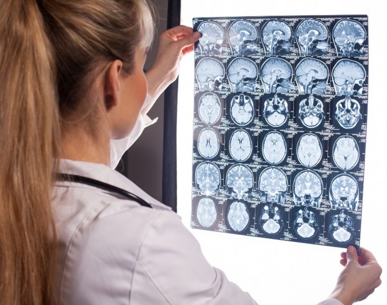
MRI scans can help to detect any abnormalities often associated with mild cognitive impairment, which can then be used to predict if you may develop dementia most commonly Alzheimers.;
They will be able to measure the size, and number, of cells in the hippocampus an area of the brain that is responsible for accessing memories.;
Often, this is the first noticeable brain function to be impaired by dementia.;
A healthy brain cortex should appear wrinkled with tissue ridges throughout, and include valleys that separate them.;
But, in brains where cortical atrophy is occurring, these ridges will appear thinner and the valleys wider.;
As dementia develops, MRIs will begin to identify changes in the brains structure, showing a decrease in the size of different parts such as the temporal and parietal lobes.;
The parietal lobe handles a number of integral functions such as calculation, order, the bodys sense of location and visual perception.;
An MRI can also demonstrate if this area of the brain has begun to atrophy, again indicating the progression of dementia.
Can An Mri Detect Alzheimers Disease
By Darlene Ortiz 8 am on February 16, 2015
Alzheimers disease , which ranks as the fourth most common cause of death in the United States, can be difficult to definitively diagnose. The disease, which is characterized by the progressive onset of dementia, causes an individual to exhibit a number of symptoms ranging from confusion and forgetfulness to changes in mood and behavior.
As a leading provider of Alzheimers care in Jefferson County, were always looking to share some of the new and innovative diagnosis and treatment options in regards to memory care. Today, were going share some insight into using MRIs to detect Alzheimers disease its early stages doing so can help to enhance future health and quality of life for seniors and aging adults.
There is no single method used to prove that an individual has Alzheimers. Although a complete assessment usually includes several techniques such as genetic testing, neurological screening and a mental status exam brain imaging techniques like MRI and CT are playing an increasingly important part in the medical communitys efforts to better understand and diagnose Alzheimers.
For more information about Alzheimers care or to learn more about caring for a senior adult with a memory condition, visit our website at www.homecareassistancejeffersonco.com or contact us directly at 303-987-5992 and schedule a complimentary, no-obligation consultation.
Don’t Miss: Does Medicare Cover Nursing Home Care For Dementia
Does An Mri Or Ct Scan Show Dementia
Ask U.S. doctors your own question and get educational, text answers â it’s anonymous and free!
Ask U.S. doctors your own question and get educational, text answers â it’s anonymous and free!
HealthTap doctors are based in the U.S., board certified, and available by text or video.
Why Early Detection Can Be Difficult

Alzheimers disease usually is not diagnosed in the early stages, even in people who visit their primary care doctors with memory complaints.
- People and their families generally underreport the symptoms.
- They may confuse them with normal signs of aging.
- The symptoms may emerge so gradually that the person affected doesnt recognize them.
- The person may be aware of some symptoms but go to great lengths to conceal them.
Recognizing symptoms early is crucial because medication to control symptoms is most effective in the early stages of the disease and early diagnosis allows the individual and his or her family members to plan for the future. If you or a loved one is experiencing any of the following symptoms, contact a physician.
Also Check: Does Alzheimer’s Run In Families
Can An Mri Diagnose Alzheimers
The simplest answer to the question is yes. The more complicated answer considers that there is still a lot of research to do on this disease, so it may be a while before we establish a definitive test to diagnose Alzheimers disease.
However, for the time being, using an MRI to detect Alzheimers is one of the best options available.
The Mri Scan: How Can It Be Used In The Diagnosis Of Alzheimer’s
As medicine has improved in the last few decades, the role of these;brain imaging studies;has become more and more important in the diagnosis of the Alzheimer’s Disease and other neurological conditions. One of the key diagnostic tests is known as Magnetic resonance imaging; the MRI.
In laymen’s terms, an MRI scan;is an imaging study that relies on a magnetic field in order to get a good view of your internal organs. The magnetic field can actually influence the water molecules that are naturally found in your body in order to give a very refined picture of what is going on in your body. It is best to use this type of imaging for soft tissue; the exact type of tissue that you will be looking at when you are investigating the brain.;
An MRI scan;can be a rather long examination for patients to endure and patients will need to lie as still as possible in order for doctors to get an accurate picture of the structures being investigated. During just a simple head investigation, patients may be asked to lie still for at least 30 minutes. This may sound like a simple task but the tubing in the room is rather small so many patients may not enjoy the slight claustrophobia;that can easily;occur. It is one of the best studies to do, though,;so the accurate results are;worth the slight discomfort for the patient.;
Read Also: How Fast Does Dementia Kill
Utility Of Fdg Pet In The Study Of Ad
The Pattern of FDG Hypometabolism Is an Endophenotype of AD
A substantial body of work over many years has identified a FDG-PET endophenotype of AD that is, a characteristic or signature ensemble of limbic and association regions that are typically hypometabolic in clinically established AD patients . The anatomy of the AD signature includes posterior midline cortices of the parietal and posterior cingulate gyri, the inferior parietal lobule, posterolateral portions of the temporal lobe, as well as the hippocampus and medial temporal cortices. Metabolic deficits in AD gradually worsen throughout the course of the disease. Bilateral asymmetry is common at early stages, more advanced disease usually involves prefrontal association areas, and in due course even primary cortices may be affected. Interestingly, the regions initially hypometabolic in AD are anatomically and functionally interconnected and form part of the large-scale distributed brain network known as the default mode network . We now know in addition that these regions are highly vulnerable to amyloid- deposition .
FDG Hypometabolism Is Related to Other AD Biomarkers and to Genes
FDG PET Is a Valid AD Biomarker
When Doctors Prescribe Brain Scans
Your physician might suggest that you get a brain scan to identify underlying problems causing mental conditions or affecting your general wellbeing.
Typically they are used to detect tumors, strokes, as well as other problems THAT CAN spark dementia that may appear on brain scans.
The cortex of the brain appears overly wrinkled and it has gyri which are separated by sulci .
Individuals with cortical atrophy experience the progressive loss of neurons which in turn causes the thinning of the ridges and the sulci to grow wider.
When brain cells continue dying, the brains fluid-filled cavities expand and occupy the available space.
In turn, they become LARGER than normal.
These structural changes within the brain are also aspects that BRAIN SCANS CAN IDENTIFY.
Also Check: What Is The Difference Between Dementia And Senility
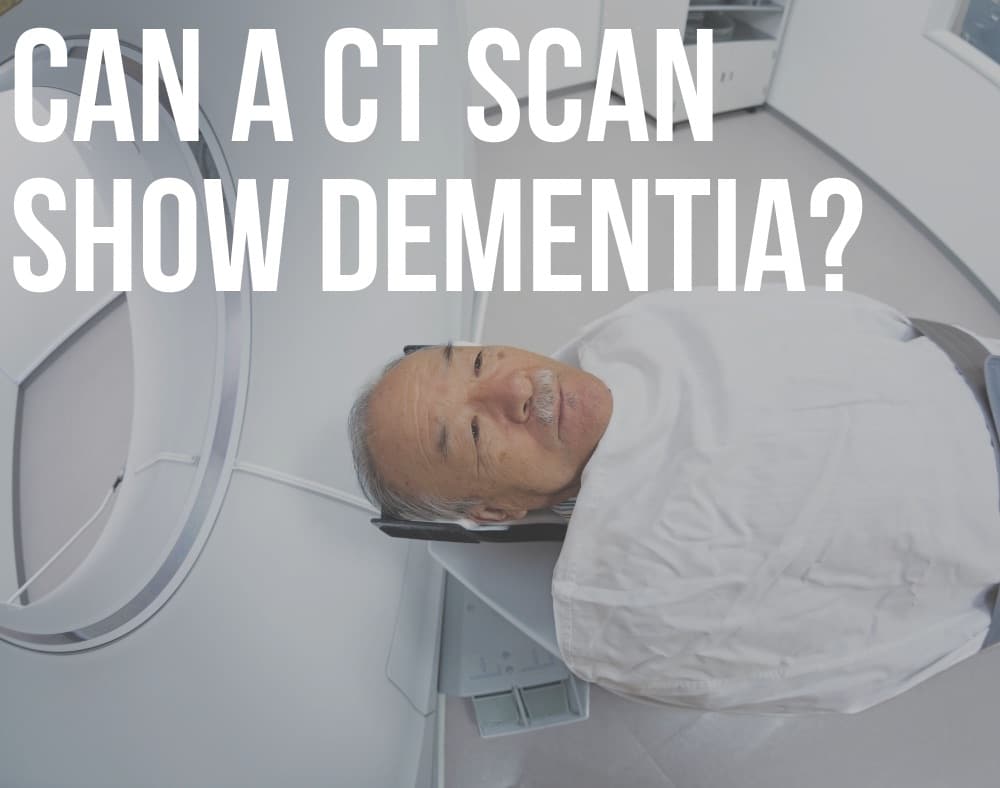
How Do Ct Scans Show Dementia
The most common types of brain scan you might encounter are magnetic resonance imaging and computed tomographic scans.
Doctors regularly recommend MRIs and CT scans when they examine someone they suspect has dementia.
CT scans detect brain structures through X-rays and the procedure can reveal evidence of ischemia, brain atrophy, and strokes.
The procedure also picks up on PROBLEMS like subdural hematomas, hydrocephalus, and changes that affect the blood vessels.
As implied, MRIs make use of focused radio waves and magnetic fields to detect the presence of hydrogen atoms within the bodys tissues.
MRIs ARE BETTER at diagnosing brain atrophy and the damage that subtle ischemia or incidents of small strokes cause to the brain.
Thus, MRI is normally the first test a person undergoes and CT second.
Utility Of Amyloid Pet In The Study Of Ad
The obvious strength of amyloid imaging is that it has allowed the determination of brain A content to be moved from the pathology laboratory into the clinic. Amyloid imaging can detect cerebral -amyloidosis and appears specific for this type of amyloid pathology, giving negative signals in pathologically confirmed cases of prion amyloid , pathologically confirmed pure -synucleinopathy , as well as in apparently pure cases of tauopathy in semantic dementia .
PiB PET Images of normal control, MCI, and AD subjects showing a range of amyloid- deposition. Most controls show no evidence of amyloid- deposition , but a substantial portion do . Most patients with MCI show moderate or severe amyloid- deposition , but as many as 40%50% show no evidence of amyloid- pathology . The vast majority of clinically diagnosed AD patients show heavy amyloid- deposition .
In the setting of MCI, combined data from nine amyloid PET studies show that 161 of 272 MCI patients were amyloid positive . Five of these studies included longitudinal clinical follow-up for 13 years on 155 MCI patients and showed that 57 of these 155 progressed to clinical AD and 53 of these 57 were amyloid positive at baseline ; only four of 54 amyloid-negative MCI patients progressed to clinical AD in these studies .
You May Like: Senile Dementia Of The Alzheimer Type
Other Imaging Options That Can Diagnose Dementia
Several other brain imaging procedures exist. Each can help detect dementia in different ways.
EEGs
EEGs are sometimes used on people who have suspected seizures, which accompany some types of dementia.
The procedure involves placing several electrodes at different points on the scalp to check for abnormalities in the brain through the recorded patterns of electrical activity.
The electrical activity shows instances of cognitive dysfunction that plague parts of the brain or the entire organ.
People with MODERATE to SEVERE cases of dementia present abnormal EEGs.
The procedure can also identify seizures, which 10% of people with Alzheimers are reported to experience.
Functional Brain Imaging
Functional brain imaging procedures are not often used as diagnostic tools. But they help researchers in the process of studying people with dementia.
They include functional single-photon emission computed tomography , MRI , magnetoencephalography , and positron emission tomography scans.
Nowadays, they have a hand in the EARLY DETECTION of dementia.
fMRI measures metabolic changes happening within the brain using strong magnetic fields.
SPECT scans reveal blood distribution within the brain. This aspect is responsible for discovering increased brain activity.
PET scans pick up on blood flow, glucose, and oxygen metabolism, and if amyloid proteins are present within the brain.
MEG scans record the electromagnetic fields that the brain produces through neuronal activities.
Pet Imaging May Also Be Used While Diagnosing Alzheimer’s

Positron-Emissions Tomography, or the PET scan, is the last type of investigation that we will spotlight in this article. There is no need to get into the complicated physics of what is happening to explain how this instrument works, but the simple version may be helpful. A PET scan;is a type of study that is used to determine how much glucose is being used by tissue. This can give a reasonable representation of how quickly the brain is metabolizing the sugar. The more active an area, the more glucose will be needed in order to maintain these functions.;
The application of this PET scan in the diagnosis of Alzheimer’s Disease is apparent in modern medicine. There are numerous studies that have shown the PET scan to be a very good tool in diagnosing the early changes in the brain. It is also able to differentiate more precisely between normal aging and patterns likely seen only in patients who have Alzheimer’s Disease, making it a fairly accurate diagnostic tool. Another advantage of this type of investigation is that it can produce models that can predict the speed of memory decline in the future. This can help doctors target specific treatments and identify symptoms that may become more pronounced depending on the region of the brain involved in the patient.;
You May Like: Senile Dementia Of Alzheimer’s Type
What Are The Benefits Of Early Diagnosis
Early planning and assistanceEarly diagnosis enables a person with dementia and their family to receive help in understanding and adjusting to the diagnosis and to prepare for the future in an appropriate way. This might include making legal and financial arrangements, changes to living arrangements, and finding out about aids and services that will enhance quality of life for people with dementia and their family and friends. Early diagnosis can allow the individual to have an active role in decision making and planning for the future while families can educate themselves about the disease and learn effective ways of interacting with the person with dementia.
Checking concernsChanges in memory and thinking ability can be very worrying. Symptoms of dementia can be caused by several different diseases and conditions, some of which are treatable and reversible, including infections, depression, medication side-effects or nutritional deficiencies. The sooner the cause of dementia symptoms is identified, the sooner treatment can begin. Asking a doctor to check any symptoms and to identify the cause of symptoms can bring relief to people and their families.
Understanding The Biology Of Ad
Importantly, imaging has a major role to play in improving our understanding of this disease . Uniquely, imaging is able to delineate in life the location within the brain of the effects of AD. Together with this topographical information imaging can quantify multiple different aspects of AD pathology and assess how they relate to each other and how they change over time. The clinical correlations of these changes and their relationships to other biomarkers and to prognosis can be studied. Ultimately the role of imaging in improving our understanding of the biology of AD underpins all its applications and is a theme that runs through the following sections of this article.
You May Like: What Is The Difference Between Dementia And Senility
How Are Mris Used In The Diagnosis Of Alzheimers Disease
The 3D imaging that MRIs for Alzheimers provide makes it easy for physicians to spot abnormalities in the brain. These abnormalities can be potential markers of Alzheimers, but they could also be symptoms of other cognitive conditions some of which may even be reversible if caught in time.
One of the first areas of the brain to be affected by Alzheimers disease;is the hippocampus, which is responsible for developing new memories and helps your brain store them. When the hippocampus shrinks as a result of Alzheimers, the brains ability to form and recall memories diminishes. An Alzheimers MRI produces 3D imaging of the hippocampus, clearly showing how many cells are present and how big it is.
The parietal lobe is another part of the brain that is negatively affected by Alzheimers. Its responsible for a lot of things, such as sensing the bodys location, doing calculations and visual perception. When the parietal lobe shrinks, it reduces our ability to do any of those things. The parietal lobes size can also indicate to doctors whether the patient is experiencing an onset of Alzheimers or another cognitive condition.
What Is Alzheimers Disease
Alzheimers is;thought to be the result;of beta-amyloid plaques, which are thick protein deposits present in the brain, and neurofibrillary tangles abnormal structures, which form in neurons building up in the brain. This buildup causes neurons in the brain to cease working, losing connection with other neurons before dying. However, the exact cause of Alzheimers is still unknown. The condition is the most common form of dementia and can quickly progress from being mild to severe.
There are no known causes for Alzheimers since the disease is still being studied. A persons chances of developing Alzheimers tend to increase with age, but people;in their 40s and 50s;can begin showing symptoms of early-onset Alzheimers. People who have;a relative with the condition;may also be at higher risk of developing it themselves since the disease has hereditary factors.
A persons overall physical health may also be a factor in whether they develop Alzheimers disease. One study showed that people who have heart disease or other cardiovascular issues could be at a higher risk of developing Alzheimers;than those with good heart health. This is because cardiovascular issues often reduce the amount of blood the brain receives, which may increase the cognitive issues associated with Alzheimers.
You May Like: Senile Dementia Of The Alzheimer Type