About Dementia And Ptsd
Dementia is relatively common in older adults, affecting about one in six women and one in ten men, according to the Institute for Dementia Research & Prevention at Pennington Biomedical Research Center at Louisiana State University System.
An estimated 7.8 percent of people in the United States will experience PTSD at some point in life, according to the Nebraska Department of Veterans’ Affairs. Approximately 30 percent of people who have spent time in a war zone will develop PTSD.
Dementia is not a specific disease, according to the Alzheimer’s Association, but rather an umbrella term that describes a wide range of symptoms.
The human brain has many distinct regions, and each is responsible for carrying out different functions, such as memory, judgment or movement. When brain cells within a certain region sustain damage, the brain cells in that region have trouble performing their appointed tasks. Damage to cells in the region responsible for memory causes memory problems, for example. In other words, the symptoms correspond to the area of the brain affected by cell damage.
Dementia is the result of damage to the brain cells. The damage prevents the brain cells from communicating with each other in ways that affect how a person thinks, feels, and behaves.
Not all types of dementia cause tangles and plaque, though. Research shows that MRI can help doctors differentiate between Alzheimer’s disease and other types of dementia.;
Oxford Brain Diagnostics: Turning Mri Into A Diagnosis Tool For Dementia
A brain reconstruction from an MRI scan using Oxford Brain Diagnostics software allows researchers to explore neurodegenerative changes.Credit: Oxford Brain Diagnostics
Oxford Brain Diagnostics is a spin-off from the University of Oxford, UK.
Neuroscientist Steven Chance spent 20 years looking at brain tissue through microscopes and examining magnetic resonance imaging scans. During this time, he became increasingly interested in the information gap between the two. Histological analysis using microscopes reveals cellular scale changes, but only after death. MRI, by contrast, is safe for use in people, but can visualize only relatively large-scale changes. Chance wanted the best of both worlds: an index of cellular scale changes, in life, he says. Thats the trajectory I was on. Such changes reveal the neurodegeneration underlying numerous forms of dementia, including Alzheimers disease. There are still no therapies that alter these processes, and the World Health Organization estimates that 152 million people worldwide will be living with dementia by 2050.
In 2010, Chance teamed up with Mark Jenkinson, a neuroimaging researcher at the University of Oxfords Wellcome Centre for Integrative Neuroimaging, UK. Eight years later, they founded Oxford Brain Diagnostics, with the aim of using specialized brain-imaging analysis to diagnose neurodegenerative conditions earlier, differentiate between them better and guide treatment decisions.
Dementia And Ptsd Diagnosis: How A 3t Mri Could Help
Mental health issues, such as dementia and post-traumatic stress disorder , can significantly decrease the quality of life in the patients who suffer them. In many cases, the signs and symptoms of these mental health issues are quite similar; this can lead to incomplete or even inaccurate diagnoses. There is an association between dementia and PTSD with the presence of one condition increasing the risk of the other and this close association can make it difficult for doctors to diagnose patients with these common mental health issues. Fortunately, advanced imaging can help.;
Read Also: What Is Vascular Dementia Life Expectancy
How Do Ct Scans Show Dementia
The most common types of brain scan you might encounter are magnetic resonance imaging and computed tomographic scans.
Doctors regularly recommend MRIs and CT scans when they examine someone they suspect has dementia.
CT scans detect brain structures through X-rays and the procedure can reveal evidence of ischemia, brain atrophy, and strokes.
The procedure also picks up on PROBLEMS like subdural hematomas, hydrocephalus, and changes that affect the blood vessels.
As implied, MRIs make use of focused radio waves and magnetic fields to detect the presence of hydrogen atoms within the bodys tissues.
MRIs ARE BETTER at diagnosing brain atrophy and the damage that subtle ischemia or incidents of small strokes cause to the brain.
Thus, MRI is normally the first test a person undergoes and CT second.
Early Warning Signs And Diagnosis

Alzheimers Disease can be caught in the early stageswhen the best treatments are availableby watching for telltale warning signs. If you recognize the warning signs in yourself or a loved one, make an appointment to see your physician right away. Brain imaging technology can diagnose Alzheimers early, improving the opportunities for symptom management.
Read Also: What Is The Difference Between Dementia And Senility
Strategic Infarcts And Small Vessel Disease
Cognitive dysfunction in VaD can be the result of :
- Large vessel infarctions:
- Bilateral in the anterior cerebral artery territory.
- Parietotemporal- and temporo-occipital association areas of the dominant hemisphere
- oPosterior cerebral artery territory infarction of the paramedian thalamic region and inferior medial temporal lobe of the dominant hemisphere
- Multiple lacunar infactions in frontal white matter and basal ganglia
- WMLs
- Bilateral thalamic lesions
MTA in a patient with VaD
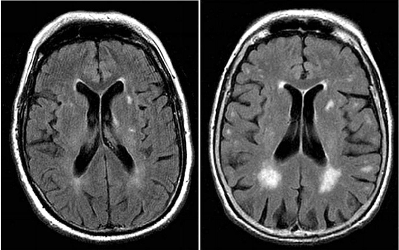
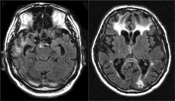
There is an increasing awareness for the importance of small vessel disease as a predictor of cognitive decline and dementia. Moreover, it seems to amplify the effects of pathologic changes of Alzheimer’s disease.On the left we see a patient who was diagnosed as having VaD.White matter disease is seen as severe WMH in the periventricular regions.In addition to these vascular changes, there is also MTA.Presumably this patient has both VaD and AD, a finding seen in many elderly patients. These findings should be described separately as it may have therapeutic consequences.
Bilateral medial strategic thalamus infarctions
The medial nuclei of the thalamus play an important role in memory and learning. A large unilateral infarction or bilateral infarctions in this region can cause dementia.You have to pay special attention to these areas to find these small infarctions.
FLAIR misses thalamus infarctionsCerebral Amyloid Angiopathy
How A Head Ct Scan Can Detect Alzheimers Disease
A head CT scan looks at the structure of your brain. This scan can detect issues such as tumors, hemorrhages, and strokes, which can all mimic the symptoms of Alzheimers, but in addition to helping you rule out those conditions, a CT scan can also detect the loss of brain mass thats associated with Alzheimers disease.
You May Like: Scientists Say Smelling Farts Prevents Cancer
Koedam Score For Parietal Atrophy
In addition to medial temporal lobe atrophy, parietal atrophy also has a positive predictive value in the diagnosis of AD. Atrophy of the precuneus is particularly characteristic of AD . This is particularly the case in young patients with AD , who may have normal MTA-scores.The Koedam scale rates parietal atrophy – assessed in sagittal, coronal and axial planes. In these planes, widening of the posterior cingulate and parieto-occipital sulci as well as parietal atrophy is rated .
Koedam scale grade 0-1
Sagittal T1-, axial FLAIR- and coronal T1-weighted images illustrating the Koedam scale of posterior atrophy. When different scores are obtained in different orientations, the highest score must be considered .
Koedam scale grade 2-3
Koedam scale grade 2-3Sagittal T1-, axial FLAIR- and coronal T1-weighted images illustrating the Koedam scale of posterior atrophy. The yellow arrows point to extreme widening of the posterior cingulate en parieto-occipital sulci in a patient with grade 3 posterior atrophy.
Mri May Help Doctors Differentiate Causes Of Memory Loss
UCLA Health
Using a software program, UCLA Health researchers were able to measure the volume of different regions of the brain, identifying areas where shrinkage may have occurred.
A UCLA-led study has found that MRI scans can help doctors distinguish whether a persons memory loss is being caused by Alzheimers disease or by traumatic brain injury.
The study, which also involved researchers at Washington University in St. Louis, is important because it could help prevent doctors from misdiagnosing Alzheimers disease a diagnosis that can be devastating for patients and their families, and can prevent them from receiving appropriate treatment. 30831-7/abstract” rel=”nofollow”>A 2016 study by researchers affiliated with the University of Toronto found that up to 21 percent of older adults with dementia may be misdiagnosed with Alzheimers.)
The current study, published in the Journal of Alzheimers disease, involved 40 patients whose average age was just under 68 and who were being treated by UCLA neurologists. All of the patients had suffered traumatic brain injury and later developed memory problems.
Dr. Cyrus Raji, the studys corresponding author and an assistant professor of radiology at Washington University, said one of the benefits of the approach is that it doesnt require specialized equipment beyond an MRI machine and the software the researchers used so it could potentially be performed at many medical centers.
Also Check: What Is The Color For Dementia
How Does An Mri Work
An MRI scan works by using a powerful magnet, radio waves, and a computer to create detailed images. Your body is made up of millions of hydrogen atoms , which are magnetic. When your body is placed in the magnetic field, these atoms align with the field, much like a compass points to the North Pole. A radio wave “knocks down” the atoms and disrupts their polarity. The sensor detects the time it takes for the atoms to return to their original alignment. In essence, MRI measures the water content of different tissues, which is processed by the computer to create a black and white image. The image is highly detailed and can show even the smallest abnormality.
Similar to CT, MRI allows your doctor to see your body in narrow slices, each about one quarter of an inch thick. For example, imagine that you are slicing a loaf of bread and taking a picture of each slice. It can view slices from the bottom , front , or sides , depending on what your doctor needs to see.
A dye may be injected into your bloodstream to enhance certain tissues. The dye contains gadolinium, which has magnetic properties. It circulates through the blood stream and is absorbed in certain tissues, which then stand out on the scan.
Selection Of Patients With Dementia
Eighty patients over the age of 60 years who fulfilled DSM IV criteria for dementia were recruited. Seventy five patients were obtained from a community dwelling population of patients referred to local old age psychiatry and geriatric medicine services for evaluation of possible dementia. Five patients were recruited from a dementia research clinic. The research was approved by the local ethics committee and all patients, as well as their nearest relative, gave informed consent.
Also Check: Ribbon Color For Dementia
What Does An Mri Do
MRI scans can help to detect any abnormalities often associated with mild cognitive impairment, which can then be used to predict if you may develop dementia most commonly Alzheimers.;
They will be able to measure the size, and number, of cells in the hippocampus an area of the brain that is responsible for accessing memories.;
Often, this is the first noticeable brain function to be impaired by dementia.;
A healthy brain cortex should appear wrinkled with tissue ridges throughout, and include valleys that separate them.;
But, in brains where cortical atrophy is occurring, these ridges will appear thinner and the valleys wider.;
As dementia develops, MRIs will begin to identify changes in the brains structure, showing a decrease in the size of different parts such as the temporal and parietal lobes.;
The parietal lobe handles a number of integral functions such as calculation, order, the bodys sense of location and visual perception.;
An MRI can also demonstrate if this area of the brain has begun to atrophy, again indicating the progression of dementia.
Additional Tests To Treat And Manage Dementia

Once an individual is diagnosed with dementia, the next step that follows is helping them understand how the condition will affect them and how to manage it.
Several other medical assessments exist to help physicians understand how the condition affects a specific person. And also help families, as well as caregivers, figure out the best course of treatment for the individual.
Did you HEAR of the peanut butter test?
Neuropsychological Testing
Neuropsychologists and psychologists who have specialized training can also prescribe neuropsychological tests to detect dementia.
The process involves WRITTEN and ORAL tests that can take several hours to complete.
They use these methods to assess the cognitive functions of the person suspected to have dementia.
It helps them figure out if certain areas are impaired.
The tests assess aspects like vision-motor, memory, comprehension, reasoning, coordination, and writing abilities.
Physicians may administer additional tests to find out if the person in question is SUFFERING from mood problems or dementia.
Functional Assessments
Dementia is a cognitive disorder that affects the afflicted persons daily functioning in different regards.
Objective assessments can establish what a person is STILL ABLE to do versus what they can no longer do in light of the condition.
Family members are asked to fillquestionnaires that provide details about the persons daily life in terms of the activities they are able to perform.
Psychosocial evaluation
Don’t Miss: Parkinsons And Alzheimers Together
Does An Mri Or Ct Scan Show Dementia
Ask U.S. doctors your own question and get educational, text answers â it’s anonymous and free!
Ask U.S. doctors your own question and get educational, text answers â it’s anonymous and free!
HealthTap doctors are based in the U.S., board certified, and available by text or video.
Why Doctors Consider Mri To Detect Dementia
Medical experts will advise on the use of MRI when they suspect that a person has dementia.
MRI uses focused radio waves and magnetic fields to detect the presence of hydrogen atoms in tissues in the human body.
MRI scans also reveal the brains anatomic structure with 3D imaging allowing doctors to get a clear view of the current state of the organ.
This way, the doctor is able to rule out other health problems like hydrocephalus, hemorrhage, stroke, and tumors that can mimic dementia.
With these scans, physicians can also detect loss of brain mass that relates to different types of dementia.
fMRI records blood flow changes that are linked to the activities of the brain. This may help physicians differentiate dementia types.
Verywellhealth.com also suggests that MRI scans can at times identify reversible cognitive decline.
In such a case, a doctor will recommend appropriate treatment that will reverse this decline and restore cognitive functioning.
You May Like: Does Meredith Have Alzheimer’s
Why Early Detection Can Be Difficult
Alzheimers disease usually is not diagnosed in the early stages, even in people who visit their primary care doctors with memory complaints.
- People and their families generally underreport the symptoms.
- They may confuse them with normal signs of aging.
- The symptoms may emerge so gradually that the person affected doesnt recognize them.
- The person may be aware of some symptoms but go to great lengths to conceal them.
Recognizing symptoms early is crucial because medication to control symptoms is most effective in the early stages of the disease and early diagnosis allows the individual and his or her family members to plan for the future. If you or a loved one is experiencing any of the following symptoms, contact a physician.
Schedule An Mri For Alzheimers Today
Early diagnosis is critical to slowing the progression of Alzheimers, and;an MRI of the head;is one of the best ways to do it. At Envision Imaging, were dedicated to providing world-class diagnostic imaging to enhance the quality of life for our patients.
No matter which of;our many locations;you visit, youll receive only the very best service from our staff of professionals who understand the stress that can surround a persons visit, so we ensure each client gets focused service with an excellent quality of care.
Find a;location near you;to schedule your MRI appointment today.
Also Check: Dementia Ribbon Color
Can An Mri Diagnose Alzheimers
The simplest answer to the question is yes. The more complicated answer considers that there is still a lot of research to do on this disease, so it may be a while before we establish a definitive test to diagnose Alzheimers disease.
However, for the time being, using an MRI to detect Alzheimers is one of the best options available.
Choose A Provider Who Thinks Skills Not Just Pills
Finding a psychotherapist or psychiatrist who can give you the tools you need, rather than simply giving you a prescription for medication can help you in the long run. Medications can be an important part of a treatment plan, but they should not be the first and only option recommended.
Amen Clinics has built the worlds largest database of functional brain scans related to emotional, behavioral, and learning issues and utilizes brain SPECT imaging as part of a comprehensive evaluation. SPECT scans allow us to more accurately diagnose and more effectively treat our patients.
Learn more about how a brain scan and a personalized treatment program that includes optimizing your biological, psychological, social, and spiritual factors can help you overcome cognitive issues. Find out how we can help you by calling 888-288-9834 or schedule a visit.
You May Like: What Is The Color For Dementia
What Are The Risks
MRI is very safe. There are no known health risks associated with the magnetic field or the radio waves used by the machine. Some people are sensitive to the contrast agent and may develop an allergic reaction. All contrast agents are FDA-approved and safe.
Be sure to tell your doctor if you have diabetes or kidney problems. In some cases a kidney function test may be needed prior to the MRI to make sure your kidneys are able to clear the contrast agent from your body.
Some special circumstances limit the use of a magnetic field, so itâs important for you to tell your doctor if any of the following apply to you:
- cardiac pacemaker or artificial heart valve
- metal plate, pin, or other metallic implant
- piercings
- intrauterine device, such as Copper-7 IUD
- insulin or other drug pump
- aneurysm clips
- cochlear implant or other hearing device
- employment history as a metalworker
- permanent eye-liner
Any metallic substance on your body can affect the quality of the images. It can also cause discomfort or injury to you when placed in the magnetic field, and may exclude you from the exam.
Also, be sure to tell your doctor if youâre pregnant. The American College of Radiology recommends that MRI scanning not be done in the first trimester of pregnancy. After the first trimester, there is no definitive research indicating that MRI is contraindicated in pregnancy. However, you will need to obtain a written order from your gynecologist for the test to be performed.