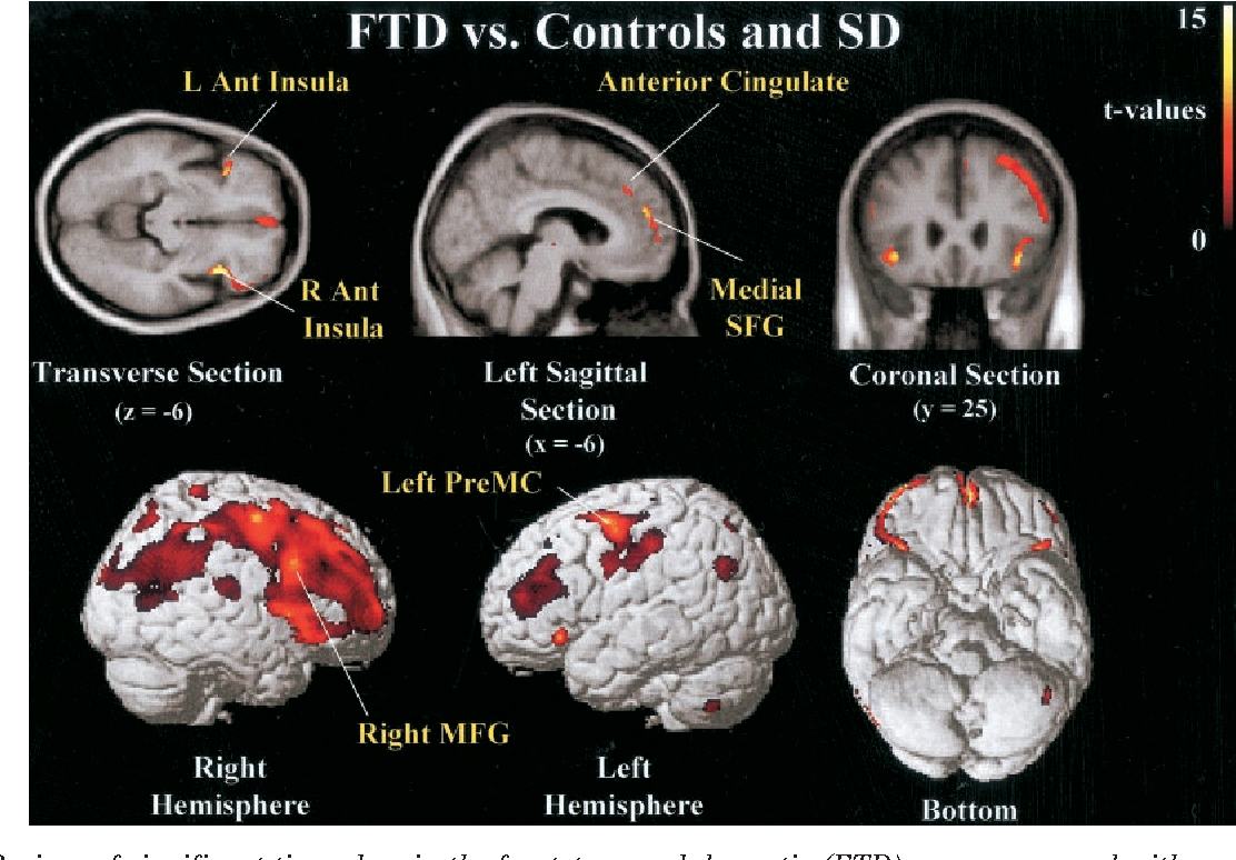
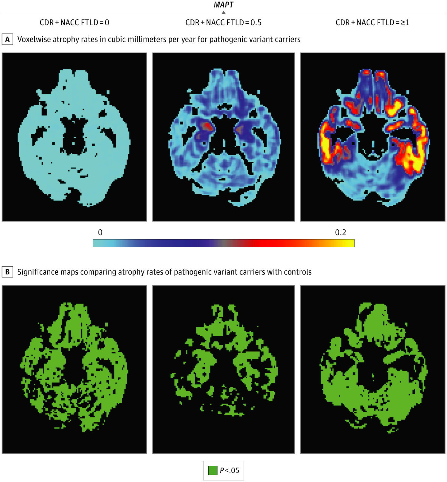
Causes Of Dementia With Lewy Bodies
Lewy bodies are tiny clumps of a protein called alpha-synuclein that can develop inside brain cells.
These clumps damage the way the cells work and communicate with each other, and the brain cells eventually die.
Dementia with Lewy bodies is closely related to Parkinson’s disease and often has some of the same symptoms, including difficulty with movement and a higher risk of falls.
Read more about dementia with Lewy bodies.
Brain Atrophy In Hiv + Patients
Brain atrophy is one of the most common neuropathologic findings in patients infected with HIV. Both head CT and structural brain MRI can demonstrate brain atrophy . CT was used to evaluate HIV-associated brain atrophy, primarily in clinical settings, over the last three decades. Studies in perinatally HIV-infected children using CT scan often showed cerebral atrophy, basal ganglia calcification, and ventricular dilatation . Early CT studies demonstrated strong interrelationships between progressive caudate atrophy and HIV-associated dementia . In patients with AIDS dementia complex, CT typically showed diffuse, symmetric cerebral atrophy that was greater than expected for the HIV patients age .
Table 18.2. Neuroimaging studies of human immunodeficiency virus brain infection
| Pathology |
|---|
|
Causes Of Brain Atrophy
Perhaps the most common question surrounding brain atrophy would be: What causes it? A number of reasons can come into play, among them normal aging.
As we age, we lose brain cells and their connections at a rate faster than we can make new cells or new connections . In fact, from young adulthood onwards, the average brain shrinks 1.9 percent in every 10-year period. In healthy people, the effects may become noticeable in their 60s, when the rate of loss increases to around 1 percent each year. The hippocampusthe area of the brain responsible for forming new memoriesshrinks significantly.
A healthy lifestyleincluding a nutritious diet, regular exercise, mental stimulation, adequate sleep, and social interactioncan slow progression of symptoms due to this normal aging process.
Also Check: How To Get A Person With Dementia To Shower
Rarer Causes Of Dementia
There are many rarer diseases and conditions that can lead to dementia, or dementia-like symptoms.
These conditions account for only 5% of dementia cases in the UK.
They include:
- problems with planning and reasoning
These symptoms are not severe enough to cause problems in everyday life.
MCI can be caused by an underlying illness, such as depression, anxiety or thyroid problems.
If the underlying illness is treated or managed, symptoms of MCI often disappear and cause no further problems.
But in some cases, people with MCI are at increased risk of going on to develop dementia, which is usually caused by Alzheimer’s disease.
Read more about how to prevent dementia.
What Are Tumors And Subdural Hematomas

Brain tumors can arise from any number of conditions or situations, including any tumor inside the cranium, or in the central spinal canal. They can be cancerous or non-cancerous in nature. Any kind of brain tumor can pose a serious risk to an individual’s health and life, due to its invasive nature.
A subdural hematoma is a clot of blood just beneath the outer covering of the brain. Usually occurring in patients over the age of 60, these clots typically form in conjunction with an atrophy of the brain.
Minor head trauma can damage the brain surface’s blood vessels, and slowly accumulate blood over several days. Most often, SDHs become very large before they are noticed because of the lack of symptoms in the early stages.
Recommended Reading: Dementia Ribbon
Harmful Alcohol Use: Imaging Studies Of Neurotoxic Effects
Brain atrophy following chronic alcohol intake is a well-known phenomenon with considerable variation across individuals. Alcohol-associated atrophy is particularly prominent in the frontal lobes however, further morphological alterations are observed such as ventricular enlargement, cerebellar atrophy, and a general widening of the cerebral sulci which exceeds comparable effects of age. Neuropathological and neuroimaging studies appear to support the hypothesis that the neurotoxic effects of alcohol can be described within the model of premature aging. Indeed, studies by Edith V. Sullivan and Adolf Pfefferbaum from the Stanford University revealed greater than expected reductions in size or blood flow in the cerebral cortex, hippocampus, cerebellum, and in the corpus callosum, when comparing older with younger alcohol-dependent patients. Further, white matter integrity can be assessed with diffusion tensor imaging , which has indicated that age-related alterations of fiber tracks in the corpus callosum are increased in alcohol-dependent patients compared to age-matched controls.
Only a few studies assessed neuropsychological correlates of brain atrophy. Frontal brain atrophy was associated with motivational deficits and with dysfunctions of the working memory. One study of Kenneth M. Adams and coworkers suggested that reduced frontal glucose utilization in alcohol patients is associated with reductions in the level of executive functioning.
Causes Of Alzheimer’s Disease
Alzheimer’s disease is the most common type of dementia.
Alzheimer’s disease is thought to be caused by the abnormal build-up of 2 proteins called amyloid and tau.
Deposits of amyloid, called plaques, build up around brain cells. Deposits of tau form “tangles” within brain cells.
Researchers do not fully understand how amyloid and tau are involved in the loss of brain cells, but research into this is continuing.
As brain cells become affected in Alzheimer’s, there’s also a decrease in chemical messengers involved in sending messages, or signals, between brain cells.
Levels of 1 neurotransmitter, acetylcholine, are particularly low in the brains of people with Alzheimer’s disease.
Medicines like donepezil increase levels of acetylcholine, and improve brain function and symptoms.
These treatments are not a cure for Alzheimer’s disease, but they do help improve symptoms.
Read more about treatments for dementia.
The symptoms that people develop depend on the areas of the brain that have been damaged by the disease.
The hippocampus is often affected early on in Alzheimer’s disease. This area of the brain is responsible for laying down new memories. That’s why memory problems are one of the earliest symptoms in Alzheimer’s.
Unusual forms of Alzheimer’s disease can start with problems with vision or with language.
Read more about Alzheimer’s disease.
Don’t Miss: Neil Diamond Alzheimer’s
Viwhat Does Atrophy Teach Us About The Pathogenesis Of Ms
Studies on brain atrophy that have emerged during the last 5 years showed that the rate of brain atrophy in patients with MS is more than fivefold higher than in age- and gender-matched controls. While the rate of atrophy varies between individual patients with MS, atrophy appears to progress at about the same rate in groups of RR-MS and SP-MS patients. This indicates that the destructive pathological process progresses inexorably over many years during the course of MS.
In most patients with RR-MS, brain atrophy progresses without obvious clinical manifestations however, the rate of brain atrophy progression predicts long-term disability status. This suggests that progressive brain tissue destruction is clinically silent in patients with MS until a threshold is surpassed, beyond which compensatory mechanisms are exhausted and disability progression ensues. This strongly supports the threshold hypothesis of disability progression in MS.
Finally, preliminary studies reviewed in the next section suggest that anti-inflammatory therapy may slow the rate of brain atrophy progression during RR-MS, but thus far no study has demonstrated a beneficial effect of anti-inflammatory therapy on atrophy progression in SP-MS. These findings are consistent with the view that atrophy progression is driven to some degree by the inflammatory process in early stages of MS.
Linda Chang, Dinesh K. Shukla, in, 2018
Dementia From Brain Conditions
If you are noticing unusual cognitive changes in a loved one, it is important to investigate all of the options, especially because some types of dementia are very treatable. Brain tumors and subdural hematomas are two brain conditions with symptoms very similar to dementia.
You May Like: Does Prevagen Work For Dementia
Prevention And Prognosis Of Cerebral Atrophy
Cerebral atrophy is not usually preventable, however, there are some things you can do to reduce your risk. These include:
- Regular exercise: This can be as simple as taking frequent walks every day. By following a regular workout regimen, you can minimize the possibility of cerebral atrophy.
- Minimizing vitamin deficiencies: Ensuring that you eat a balanced and healthy diet, particularly eating foods rich in vitamins, such as B12, will give you the best chance of preventing cerebral atrophy.
- Drinking enough water: Dehydration can lead to the increase of stress hormones and acute brain damage. Therefore, it is recommended to drink plenty of water every day to stay hydrated.
- Consuming fruits and vegetables: It is recommended to eat five servings of fruits and vegetables every day. These may include blackberries, blueberries, cranberries, cherries, strawberries, spinach, raspberries, plums, broccoli, beets, oranges, and red bell peppers. They are not only delicious to eat but are packed with vitamins and minerals as well as being rich in antioxidants.
The level of brain functioning is directly related to the area of the brain affected by cerebral atrophy. In the majority of cases of focal atrophy, fatal outcomes are not particularly common but can still cause impairment of normal functioning. Cerebral atrophy outcomes will generally vary from person to person, with advanced stages often leading to complete dementia.
Related:
Neuropsychological Testing And Cognitive Classification
The DRS-2 , a measure of global cognitive performance, has been validated as an assessment instrument for Parkinson’s disease with dementia , discriminates between Parkinson’s disease with mild cognitive impairment and Parkinson’s disease with dementia , and predicts long-term conversion to Parkinson’s disease with dementia . Cognitive categories were defined on the basis of recommended age-standardized DRS-2 scores using new robust and expanded norms : Parkinson’s disease with normal cognition Parkinson’s disease with mild cognitive impairment and Parkinson’s disease with dementia . The DRS-2 total score is constructed from five subscores: memory, attention, initiation/perseveration, construction and conceptualization.
You May Like: Purple Ribbon Alzheimers
How To Diagnose Cerebral Atrophy
The only way to determine the size of the brain is to take an image of it. Medical professionals achieve this using several advanced imaging techniques, which include:
- Magnetic Resonance Imaging Scan
- Computer Tomography Scan
- Positron Emission Tomography Scan
- Single-Photon Emission Computed Tomography
MRI is considered the most sensitive test and is the preferred method for detecting focal atrophic changes. Other characteristic features of cerebral atrophy include prominent cerebral sulci and ventriculomegaly without bulging of the third ventricular recesses. Additionally, specific conditions affecting the brain will present with their own unique areas of cerebral atrophy.
Cerebral Atrophy: Why Your Brain Is Shrinking And What To Do About It

Written byMohan GarikiparithiPublished onDecember 14, 2017
Cerebral atrophy or brain atrophy refers to the progressive loss of brain cells, called neurons, leading to decreased brain size. This phenomenon can occur to the entire brain or be focused on a singular part. The most troubling issue with cerebral atrophy is the potential for it to affect brain function, as the location of lost brain cells will potentially lead to neurological side effects.
You May Like: Ribbon Color For Dementia
Atrophy On Brain Scans
Below I show an MRI brain scan. We can see the skull and the corticospinal fluid surrounding the brain . The brain itself has two colours on this type of MRI scan a withe-ish colour and a grey-ish colour. The white-ish colour indicates the so-called white matter, which is basically the nerve fibre bundles connecting different brain regions with each other. The white matter is not as important to atrophy as they grey matter the greyish coloured brain regions. The grey matter is where most of the cells bodies of our nerve cells sit. We can see that the majority of the nerve cells can be found on the outer regions of the brain the so-called brain cortex its like a grey ribbon around the brain. Since atrophy is mostly affecting the nerve cells, it is, therefore, the brain cortex , where we can see atrophy brain changes in dementia on brain scans.
Figure showing MRI brain scan, showing the brain, skull and corticospinal fluid zoomed in insert shows a close-up of the grey matter ribbon at the brain cortex red line indicates border between grey matter and corticospinal fluid blue line indicates border between grey matter and white matter.Figure shows brain MRI scans of: a healthy aged person a person with mild dementia and a person with moderate dementia. Three red square highlights the atrophy, showing increasing gaps between brain regions caused by nerve cell loss in the grey matter. Note: The scans are three different people, not the same person over time.
How Common Is Dementia And Brain Shrinkage
Although dementia and brain shrinkage has always been common, it has become even more common among the elderly in recent history. It is not clear if this increased frequency reflects a greater awareness of the symptoms or if people simply are living longer and thus are more likely to develop dementia and brain shrinkage in their older age.
Dementia caused by neurological degenerative disease, especially Alzheimers disease, is increasing in frequency more than most other types of dementia. Some researchers suspect that as many as half of all people over 85 years old develop Alzheimers disease. Dementia associated with AIDS, which appeared to be increasing in frequency in the 1990s is now much less commonly seen, since the development of highly effect anti-retroviral therapy.
Don’t Miss: Alzheimers Awareness Symbol
Same Symptoms Different Disease
Exhibiting symptoms of dementia does not necessarily indicate dementiaââ¬âit could be another kind of brain condition, such as brain tumors and SDHs.
The loss of cognitive ability and difficulty with memory retention and recall are always causes for concern. It is important to fully understand which condition is causing these symptoms, because dementia is not always the source.
0651
Additive Effect Of Cevd And Atrophy
Further Analysis of Variance models were conducted, which showed that in the none-mild CeVD group, there was no significant difference in scores between all three atrophy groups in global cognition. In subjects with significant CeVD, the CA+MTAgroup performed significantly worse than the no atrophy group and CA group , after controlling for age, gender, education, CI status and the presence of vascular risk factors.
Domain-specific cognitive z-scores in no atrophy, CA and CA+MTA groups in different CeVD groups
Notably, there was no significant difference in the global cognitive z-score for non-significant and significant CeVD groups among subjects with CA+MTA =0.80, p=0.37), after controlling for covariates. This reflects a unidirectional additive effect of atrophy on top of CeVD, but not vice versa.
Don’t Miss: Smelling Farts Prevents Cancer
Can I Prevent Cerebral Atrophy
There is no clear-cut evidence that cerebral atrophy is preventable, but taking certain steps reduces the possibility of its early or severe onset. These include exercising regularly, regulating blood pressure, and eating a healthy diet. Foods rich in antioxidants and omega-3 fatty acids are good for your gray matter.
Dementia Symptoms And Areas Of The Brain
Knowing how different types of dementia affect the brain helps explain why someone with dementia might behave in a certain way.
Dementia and the brain
Until recently, seeing changes in the brain relied on studying the brain after the person had died. But modern brain scans may show areas of reduced activity or loss of brain tissue while the person is alive. Doctors can study these brain scans while also looking at the symptoms that the person is experiencing.
The most common types of dementia each start with shrinkage of brain tissue that may be restricted to certain parts of the brain.
Recommended Reading: Does Diet Coke Cause Memory Loss
How To Support Your Brain Health As You Age
âAs you get older, there are things you can do to support your brain health and help prevent cognitive decline.â
âGet physically active every day. Getting exercise increases blood flow to your entire body, especially your brain. Experts also believe that regular exercise can help reduce stress and depression and improve memory.
Eat healthy. Did you know that eating a heart-healthy diet also benefits your brain? Foods like fresh fruits, fish, lean meat, and skinless chicken are all good options.â
âAs you get older, itâs best to avoid overusing alcohol, as too much can lead to memory issues and confusion.
âStay mentally active. Activities like reading, playing word games, taking up a new hobby, enrolling in classes, or learning how to play an instrument are all great ways to stay mentally active. Consistent mental activity can help keep your memory and thought processing in good shape.
Stay social. Keeping up with friends and family is not only enjoyable, but it also helps ward off depression and stress. You may want to try volunteering or joining an organization so you get the satisfaction of helping people while maintaining positive social interaction.
Keep an eye on cardiovascular disease. Medical diseases like high blood pressure, high cholesterol, and diabetes can increase your risk of cognitive decline. Talk with your doctor about treatment options and how they can help.â
A few fun ways to incorporate vitamin B into your diet include:â
Show Sources
Treatments For Dementia From Brain Conditions

Treatments for brain tumors vary depending on whether they are malignant or benign. Most treatments include some kind of reductive surgery, and cancerous conditions can be treated with radiation therapy or chemotherapy.
After the brain injury, the success of treatment varies. The typical treatment, once detected, is to drill a small hole in the outer layer of the skull and drain the mass of collected blood through a catheter. Approximately 80 to 90 percent of all patients treated this way eventually recover significant portions of brain functionality.
Don’t Miss: Dementia Ribbon Tattoo