Basics Of Fdg Pet As Applied To Ad
Brain FDG PET primarily indicates synaptic activity. Because the brain relies almost exclusively on glucose as its source of energy, the glucose analog FDG is a suitable indicator of brain metabolism and, when labeled with Fluorine-18 is conveniently detected with PET. The brains energy budget is overwhelmingly devoted to the maintenance of intrinsic, resting activity, which in cortex is largely maintained by glutamaturgic synaptic signaling . FDG uptake strongly correlates at autopsy with levels of the synaptic vesicle protein synaptophysin . Hence, FDG PET is widely accepted to be a valid biomarker of overall brain metabolism to which ionic gradient maintenance for synaptic activity is the principal contributor . In this context, a single, specific AD-related alteration in FDG metabolism has not been identified and therefore the FDG-PET abnormalities described below are assumed to be the net result of some combination of processes putatively involved in the pathogenesis of AD including, but not limited to, expression of specific genes, mitochondrial dysfunction, oxidative stress, deranged plasticity, excitotoxicity, glial activation and inflammation, synapse loss, and cell death.
Early Diagnosis Of Alzheimer’s Disease And Mild Cognitive Impairment
The typical reductions of hippocampal volume in MCI with an average Mini-Mental State Exam score of 25 are 10% to 15% and in AD with an average MMSE score of 20 are 20% to 25% . Measuring these significant reductions in the medial temporal lobe can be extremely useful for early diagnosis of AD and MCI. At present, diagnostic criteria for AD are based on the criteria in the Diagnostic and Statistical Manual of Mental Disorders, Fourth Edition , which are based primarily on clinical and psychometric assessment and do not use quantitative atrophy information available in sMRI scans. However, there is a proposal to add reliable biomarkers to the diagnostic criteria . One of the suggested features is the volume loss of medial temporal structures since measures of sMRI atrophy have accuracies of 70% to 90% in AD and 50% to 70% in amnestic MCI in distinguishing them from age-matched controls . All of the above-mentioned cross-sectional methods, except 3a, can be used as diagnostic metrics for AD and MCI.
Could White Matter In Brain Mri Be A Sign Of Dementia And Ms
30, female, 230lbs, on and off cannabis smoker.
Hey, everyone. I wanted to get some opinions on an MRI that I got back in March. The MRI was because I was experiencing uneven pupils after having COVID which made me really worried.
Here were the findings of my MRI:
“There is no acute infarct, acute intracranial hemorrhage, mass lesion, mass effect, hydrocephalus, or abnormal extra-axial collection. There are a few foci of T2/FLAIR hyperintensity in the bifrontal subcortical white matter, nonspecific but of doubtful clinical significance. The dural venous sinuses maintain normal flow voids.”
The part that concerns me is the white matter. When speaking with my neurologist about my results at the time, she said that could be because of my migraines even though I told her that I wasn’t experiencing migraines. The only time I would experience headaches around this time was when I was having depressive crying that it would make my head hurt.
I was doing some research that suggests that this is usually found in older patients and can be a sign of disorders like Alzheimer’s and dementia are going to happen. Also can be signs of silent strokes.
You May Like: Can You Get Disability For Early Onset Alzheimer’s
Dementia With Lewy Bodies
Dementia with Lewy bodies is responsible for approximately 25% of dementias and belongs to the atypical Parkinson syndromes together with progressive supranuclear palsy and multi-system atrophy . The clinical manifestations can be similar to that of AD or dementia associated with Parkinson’s disease. Patients typically present with one of three symptom complexes: detailed visual hallucinations, Parkinson-like symptoms and fluctuations in alertness and attention.Pathologically, the disease is characterized by the presence of Lewy bodies in various regions of the hippocampal complex, subcortical nuclei and neocortex with a variable number of diffuse amyloid plaques. Cholinesterase inhibitors are currently the treatment of choice for this condition.
The role of imaging is limited in Lewy body dementia.Usually the MR of the brain is normal, including the hippocampus. This finding is important as it enables us to differentiate this disease from Alzheimer’ s disease, the main differential diagnosis. Nuclear imaging can be used to demonstrate an abnormal dopaminergic system
PSP with midbrain atrophy
Frederik Barkhof Marieke Hazewinkel Maja Binnewijzend And Robin Smithuis

Alzheimer Centre and Image Analysis Centre, Vrije Universiteit Medical Center, Amsterdam and the Alrijne Hospital, Leiderdorp, The Netherlands
Publicationdate 2012-01-09 / Update: 2022-03-03
This presentation will focus on the role of MRI in the diagnosis of dementia and related diseases.We will discuss the following subjects:
- Systematic assessment of MR in dementia
- MR protocol for dementia
- Typical findings in the most common dementia syndromes
- Alzheimer’s disease
Read Also: Can People With Dementia Read
Early Warning Signs And Diagnosis
Alzheimers Disease can be caught in the early stageswhen the best treatments are availableby watching for telltale warning signs. If you recognize the warning signs in yourself or a loved one, make an appointment to see your physician right away. Brain imaging technology can diagnose Alzheimers early, improving the opportunities for symptom management.
Pathological Cascade And Structural Magnetic Resonance Imaging
Alzheimer’s disease is a multifaceted disease in which cumulative pathological brain insults result in progressive cognitive decline that ultimately leads to dementia. Amyloid plaques, neurofibrillary tangles , neurodegeneration, and inflammation are the well-established pathological hallmarks of AD. A plausible model for the development of AD posits that amyloid deposition occurs early in the process but by itself does not directly cause clinical symptoms . Neuronal and synaptic losses appear to be key determinants of cognitive impairment in AD . If neuronal loss leads to cerebral atrophy , then it can be expected that cognitive decline and atrophy will be closely associated. On the basis of this evidence, it has been hypothesized that AD pathological cascade is a two-stage process in which amyloidosis and neuronal pathology are largely sequential rather than simultaneous processes . There is also sufficient literature to support the fact that atrophy of the brain structures or neurodegeneration is the most proximate substrate of cognitive impairment in AD . This hypothesis of a sequential model was proposed by Jack and colleagues on the basis of biomarker data and is adapted and illustrated in Figure . Owing to the close relationship between neurodegeneration and cognition , atrophy measured on structural magnetic resonance imaging is a powerful AD biomarker.
Figure 1
Read Also: How Many People In America Have Dementia
Single Brain Scan Can Diagnose Alzheimers Disease
by Maxine Myers20 June 2022
A single MRI scan of the brain could be enough to diagnose Alzheimers disease, according to new research by Imperial College London.
The research uses machine learning technology to look at structural features within the brain, including in regions not previously associated with Alzheimers. The advantage of the technique is its simplicity and the fact that it can identify the disease at an early stage when it can be very difficult to diagnose.
Although there is no cure for Alzheimers disease, getting a diagnosis quickly at an early stage helps patients. It allows them to access help and support, get treatment to manage their symptoms and plan for the future. Being able to accurately identify patients at an early stage of the disease will also help researchers to understand the brain changes that trigger the disease, and support development and trials of new treatments.
Comparison Of Structural Magnetic Resonance Imaging With Other Major Alzheimer’s Disease Biomarkers
The major AD biomarkers that are typically considered for clinical trials and observational studies are CSF A1-42, CSF t-tau, fluoro-deoxy-glucose positron emission tomography , Pittsburgh compound B-PET , and sMRI. In this section, we will compare sMRI with other major AD biomarkers by summarizing studies that have compared sMRI with each of these biomarkers in the same set of subjects.
Read Also: Does Insurance Cover Dementia Care
Cerebral Autosomal Dominant Arteriopathy With Subcortical Infarcts And Leukoencehalopathy
CADASIL is another hereditary disease which may present with a progressive cognitive dysfunction.Other presenting symptoms include migraines, stroke-like episodes and behavioral disturbances. It affects the small vessels of the brain.Confluent white matter hyperintesities in the frontal and especially anterior temporal lobes in combination with infarcts and microbleeds are seen on imaging.
The FLAIR images show classic findings in CADASIL – confluent white matter hyperintensities with lacunar infarcts and involvement of the anterior temporal lobes.
Traumatic brain injury
Strategic Infarcts And Small Vessel Disease
Cognitive dysfunction in VaD can be the result of :
- Large vessel infarctions:
- Bilateral in the anterior cerebral artery territory.
- Parietotemporal- and temporo-occipital association areas of the dominant hemisphere
- oPosterior cerebral artery territory infarction of the paramedian thalamic region and inferior medial temporal lobe of the dominant hemisphere
- Multiple lacunar infactions in frontal white matter and basal ganglia
- Bilateral thalamic lesions
MTA in a patient with VaD
There is an increasing awareness for the importance of small vessel disease as a predictor of cognitive decline and dementia. Moreover, it seems to amplify the effects of pathologic changes of Alzheimer’s disease.On the left we see a patient who was diagnosed as having VaD.White matter disease is seen as severe WMH in the periventricular regions.In addition to these vascular changes, there is also MTA.Presumably this patient has both VaD and AD, a finding seen in many elderly patients. These findings should be described separately as it may have therapeutic consequences.
Bilateral medial strategic thalamus infarctions
The medial nuclei of the thalamus play an important role in memory and learning. A large unilateral infarction or bilateral infarctions in this region can cause dementia.You have to pay special attention to these areas to find these small infarctions.
FLAIR misses thalamus infarctionsCerebral Amyloid Angiopathy
You May Like: How To Help Someone With Dementia Who Is In Denial
Predicting The Risk Of Progression In Mild Cognitive Impairment And Cognitively Normal
Although there is considerable variability of progression rates in MCI to AD, it has been observed that an average of about 10% to 15% of subjects with MCI, specifically of the amnestic type, annually progress to AD . Because pathological changes occur before the onset of clinical symptoms, biomarkers can aid in the prediction of risk of progression in MCI and CN. A recent meta-analysis showed that hippocampal volume can detect an average of approximately 73% of MCI subjects who progress to AD . Several studies using both cross-sectional methods 1 and 2 above have shown that atrophy seen on MRI can predict the risk of progression to AD with good accuracy.
Looking To The Future: The Role Of Imaging In The Treatment Of Ad

The search for therapies that can modify the course of ADto slow, delay, or prevent itis clearly our most important challenge. That search has in turn led to a search for imaging markers that can be used as outcomes in drug discovery and trials. The value of any imaging technology will ultimately be determined by its contribution to meeting the challenge of finding and using effective therapies. This value includes contributions toward diagnosis. The large variability, intrinsic to clinical outcomes in AD, means that studies relying purely on clinical measures are necessarily large and consequently very costly. Using clinical outcomes to power studies to establish meaningful disease-slowing effects may require complicated designs and thousands of subjects. A major aim in academia and industry has been to find biomarkers that could identify disease-slowing effects earlier and/or with significantly fewer subjects exposed to treatment. Imaging is being increasingly incorporated into trial designs to measure the effects of a therapy on fibrillary amyloid on atrophy and on metabolism .
You May Like: What Is The Longest Day For Alzheimer’s
How Is Alzheimers Disease Diagnosed
So little is known about Alzheimers that theres no single test to diagnose it. However, there are ways to test brain function, and these tests, along with examining symptoms and ruling out other potential conditions, are generally how physicians diagnose Alzheimers. Your family doctor can diagnose the disease, but psychologists, neurologists and geriatricians can also provide a diagnosis or a second opinion.
An examination for diagnosing Alzheimers disease can include:
- Complete and accurate medical history
- Test for mental status
Brain imaging allows medical professionals to see how the brain is functioning and accurately spot any abnormalities. Alzheimers imaging can include:
Changes In Brain Structure
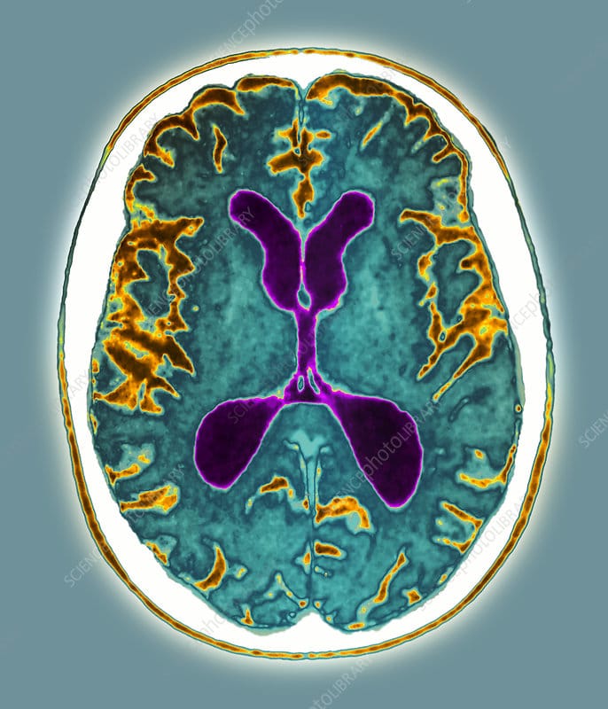
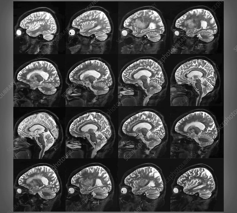
Diffuse cerebral atrophy with widened sulci and dilatation of the lateral ventricles can be observed. Disproportionate atrophy of the medial temporal lobe, particularly of the volume of the hippocampal formations , can be seen.
The hippocampus is one of the earliest affected brain regions in Alzheimer disease, and its dysfunction is believed to underlie the core feature of the disease-memory impairment. Changes in hippocampal volume, shape, symmetry, and activation are reflected by cognitive impairment.
Dilatation of the perihippocampal fissure is a useful radiologic marker for the initial diagnosis of Alzheimer disease, with a predictive accuracy of 91%. The hippocampal fissure is surrounded laterally by the hippocampus, superiorly by the dentate gyrus, and inferiorly by the subiculum. These structures are all involved in the early development of Alzheimer disease and explain the enlargement in the early stages. At the medial aspect, the fissure communicates with the ambient cistern, and its enlargement on CT scans is often seen as hippocampal lucency or hypoattenuation in the temporal area medial to the temporal horn.
The temporal horns of the lateral ventricles may be enlarged. Prominence of the choroid and hippocampal fissures and enlargement of the sylvian fissure may be noted. White matter attenuation is not a feature of Alzheimer disease.
Don’t Miss: When Does Dementia Typically Start
Utility Of Structural Mri In The Study Of Ad
Atrophy in AD
Progressive cerebral atrophy is a characteristic feature of neurodegeneration that can be visualized in life with MRI . The major contributors to atrophy are thought to be dendritic and neuronal losses. Studies of regional MRI volumes have shown these are closely related to neuronal counts at autopsy . The pattern of loss differs between diseases reflecting selective neuronal vulnerability and/or regional disease expression. AD is characterized by an insidious onset and inexorable progression of atrophy that is first manifest in the medial temporal lobe . The entorhinal cortex is typically the earliest site of atrophy, closely followed by the hippocampus, amygdala, and parahippocampus . Other structures within the limbic lobe such as the posterior cingulate are also affected early on. These losses then spread to involve the temporal neocortex and then all neocortical association areas usually in a symmetrical fashion. This sequence of progression of atrophy on MRI most closely fits histopathological studies that have derived stages for the spread of neurofibrillary tangles . Nonetheless, a significant minority of AD cases have atypical presentations and in these cases the pattern of atrophy accords with clinical phenotype: with language presentations particularly having left temporal atrophy and visual variants having posterior cortical atrophy.
Measuring Progression in AD with Structural MRI
Availability and Utility of Structural MRI
Basics Of Structural Mri As Applied To Ad
MRI utilizes the fact that protons have angular momentum which is polarized in a magnetic field. This means that a pulse of radiofrequency can alter the energy state of protons and, when the pulse is turned off, the protons will, on returning to their energy stage, emit a radiofrequency signal. By a combination of different gradients and pulses, sequences can be designed to be sensitive to different tissue characteristics. In broad terms structural MRI in AD can be divided into assessing atrophy and changes in tissue characteristics which cause signal alterations on certain sequences such as white matter hyperintensities on T2-weighted MRI as a result of vascular damage. A number of MR sequences that are sensitive to microstructural change have shown alterations in AD. These sequences are already important research tools however, they have not yet found a place in routine clinical practice in AD and they will not be considered further here.
Don’t Miss: Warning Signs Of Alzheimer’s Disease Include
Calculation Of Atrophy Rates And Sample Sizes
For a volume \ at baseline and a volume \ at a follow-up time point we calculated atrophy rates using the logarithmic transform as \=}}_^}=\)\\cdot \mathrm\). Note that atrophy rate and volume change is used interchangeably, which means that a positive atrophy rate indicates an increase in volume.
For a power and significance level the sample size can be calculated as:
Here is the difference in atrophy rate that is to be shown between the clinical groups. In this study sample sizes were calculated to detect a 25% change in atrophy rate with 80% power at a 5% significance level . These parameter choices are commonly found in the literature,,. It is important to relate atrophy rates in dementia to normal atrophy during aging, as in the uncorrected case it is assumed that 100% treatment effect would effectively reduce the structural atrophy to zero. Sample sizes were thus corrected for normal ageing by evaluating Equation with =0.25 to reduce the maximal treatment effect to the level of normal ageing. In Equation it is assumed that measurements of healthy atrophy have the same variance as measurements of diseased subjects . This usually leads to a more conservative estimate.
Differential Diagnosis Of Dementia Subtypes
Given that pathology does not always map onto the clinical expression of the disease and has considerable clinical heterogeneity, biomarkers such as sMRI can aid in the differential diagnosis of dementia types. The absence of significant medial temporal lobe atrophy in dementia with Lewy bodies and vascular dementia , significant frontal lobe atrophy in behavioral variant fronto-temporal dementia , or pronounced asymmetrical temporal lobe atrophy in semantic dementia can be used to separate these non-AD dementias from AD. Diffusion imaging and FLAIR are useful in identifying both cerebrovascular disease and prion disease. MRI is useful in identifying structural contributors to cognitive impairment such as hemorrhage or evidence of major head trauma. Differential diagnosis of dementias using sMRI will be particularly helpful when therapeutics become readily available.
Also Check: Drugs To Treat Alzheimer’s Disease