The Structure Of The Message: Grammar And Phonology
The structure of a verbal message can be considered at two levels: grammar, the ordering of words at the level of phrases and sentences, including the use of âfunction wordsâ and phonology, the selection and ordering of individual sounds into syllables and words. Impaired grammatical structure typically manifests as disjointed or âtelegraphicâ speech composed of single words and short phrases, omitting function and connecting words . Incorrect ordering of words may occur, grammatical elements such as plurals or tenses may be misused or binary grammatical alternatives may be confused . Impaired phonological structure manifests as speech sound errors, or âphonemic paraphasias’ at the level of individual words and syllables, most commonly substitutions , transpositions , omissions or additions . Such errors often first appear and remain more evident with polysyllabic words. Agrammatism and phonemic errors are typical features of PNFA and help distinguish this syndrome from the language output difficulties observed in patients with AD . Agrammatism and phonological breakdown commonly occur together but relatively pure dissociations have been described in degenerative disease . Agrammatism may be partly masked by other speech-production impairments, unless more detailed testing of the receptive aspects of sentence comprehension or written output is undertaken .
A Comparison Of Acute And Progressive Disorders With Word
Although there is considerable overlap between the disorders of word-finding in acute disease states and in the progressive aphasias, certain features are more typically seen in one setting rather than the other. These divergences are both relevant to the clinical analysis of language dysfunction in these different disease states and of considerable interest for the pathophysiological insights they provide into language neurobiology. Key clinical features of the language disturbance in selected acute and progressive disorders with prominent word-finding difficulty are summarized in the .
Functions Of The Brain
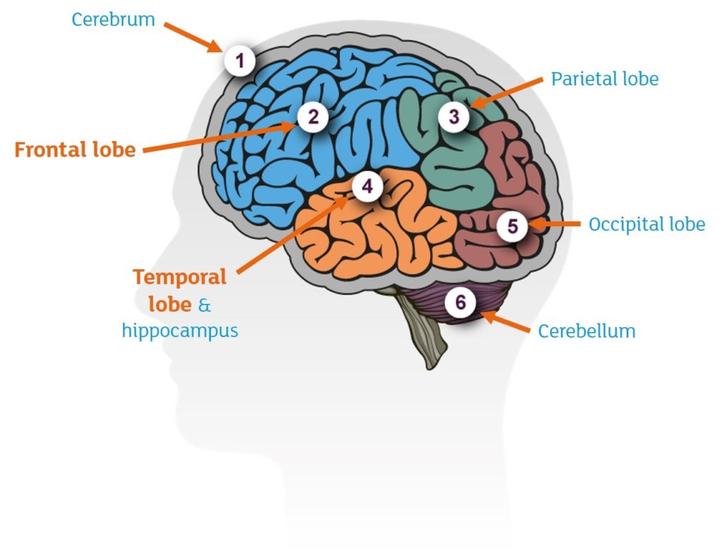
To get a good understanding of fronto-temporal dementia and semantic dementia, it is useful to look at the brain and the way it functions .
The different areas of the brain play different roles. The posterior areas are responsible for our visual functions. They help us to see what things are and where they are in space. The temporal lobes play an important role in memory and language. They enable us to learn and store new information. Finally, the frontal lobes are responsible for organisation and control of information. They enable us to generate plans and make decisions. This is often referred to as executive function
Read Also: Senile Vs Dementia
Language And Neuropsychological Testing
Other than brain imaging studies, the most specific tests for evaluating frontotemporal lobe dementia are evaluation with standardized language batteries and neuropsychological testing. Such studies assess the specific pattern of language abnormality and the presence of other cognitive and memory deficits. Preservation of many of these functions distinguishes FTD and primary progressive aphasia syndromes from Alzheimer disease.
In distinguishing FTD from Alzheimer disease, the involvement of specific cognitive functions is the most important differentiating factor.
Grossman pointed to a double dissociation between immediate and short-term memory in a comparison study of 4 patients with PPA versus 25 patients with presumed Alzheimer disease. Immediate memory was more impaired in PPA patients, whereas short-term memory deficits characterized the deficits of patients with Alzheimer disease. The frontal cortex, especially on the left side, is thought to be the site of working or immediate memory, whereas the hippocampus and other medial temporal structures, often affected early in Alzheimer disease, represent the site of short-term memory.
PPA is a syndrome, not a pathological diagnosis. Although the term initially implied a pathology other than Alzheimer disease, we must now consider that some cases may have a syndrome of PPA but a pathological diagnosis of Alzheimer disease, or vice versa.
Disinhibition
Driving And Work Problems

In 2009 Pat began having problems with her driving. In September she hit a car pulling out from a driveway and worryingly didnt stop. In December she drove through a roundabout and somehow rolled her new car over.
In 2010 problems increased at work. On July 27 she noted in her diary: J told me that my colleagues didnt have confidence in my abilities. Definitely going to resign in Dec. Sadly even working as a nurse in the NHS her employer didnt think that a rapid decline in her abilities after 35 years nursing might be due to illness.
Don’t Miss: Color For Alzheimer’s Ribbon
Life Expectancy And Treatment
About 10 15% of dementia cases are thought to be frontal lobe dementia, the disease affecting 1 in 5000 of the population. However in those under 65 it is believed to be 20 50% of cases. Onset of frontal lobe dementia is normally identified when the patient is between 45 and 65 years of age, although it has been seen in people aged 20 to 30 years of age. Only 10% of cases are identified in those 70 years and over.
The disease takes from three to ten years to progress, although there are instances of much shorter or longer times. The average life expectancy of a person diagnosed with frontal lobe dementia is eight years. Approximately 50% of deaths are as a result of pneumonia, following complications associated with inability of the person to move or care for themselves.
As with other forms of dementia there is no current cure for the disease, but there are a range of treatments that can help to manage and deal with the symptoms, and to help people to regain some of their lost functions.
These include drugs such as SSRI antidepressants to help control the symptoms like obsession, over-eating and depression. Antipsychotics may be given to address challenging and inappropriate behaviours. Psychological treatments such as cognitive stimulation and behavioural therapy can help maintain memory function address anxiety. Rehabilitative practices such as, occupational therapy, physiotherapy and speech therapy can help the brain to learn new ways to do things.
References:
Occurrence In The United States
The exact prevalence of frontotemporal lobe dementia is unknown. Among patients presenting with dementia who are younger than 65 years, the prevalence may be similar to or greater than that of Alzheimer disease. Some series based on brain pathology have estimated that FTD is responsible for as many as 10% of cases of dementia. In the United States, estimates are generally lower FTD ranks after Alzheimer disease, vascular dementia, and Lewy body dementia in frequency of dementing illnesses.
You May Like: What Color Ribbon Is Alzheimer’s
Mild Cognitive Impairment Due To Alzheimer Disease
Mild cognitive impairment consists of a heterogeneous pathology, and MCI due to AD is a transitional stage between aging and AD. MCI due to AD demonstrates the same glucose-reduction pattern as early AD , which predicts that the patient will show symptoms of AD in the near future. At the early stage of AD or MCI due to AD, it is difficult to detect the characteristic hypometabolic patterns on FDG-PET images by visual inspection. As such, statistical images are helpful. Nevertheless, FDG-PET generally has a higher accuracy than MR imaging for diagnosing early AD, and for predicting rapid conversion of MCI to early AD. A combination of PET and other biomarkers is important because imaging and CSF biomarkers can improve prediction of conversion from MCI to AD compared with baseline clinical testing. FDG -PET appears to add the greatest prognostic information.â
MCI due to AD. Regions exhibiting a significant reduction in glucose metabolism in patients with MCI due to AD compared with healthy elderly subjects are demonstrated by statistical parametric maps. Bilateral parietal and posterior cingulate metabolism is decreased in patients with MCI due to AD. These decreased regions are the same as those in patients with early AD.
The Sense Of The Message: Conceptual Content And Vocabulary
Once a plan for a verbal message is generated, the message must be elaborated with specific content and function words. The sense of a spoken thought or message depends on its conceptual content. It is possible to convey the constituent concepts of a message even where the structure is disorganized or degraded, and the converse is also true. To take the example of the message âthe bird sat on the branchâ: compare âbird sat branch or âthe birt sit on the brenchâ with âthe thing pit on the tamâ . The content of speech can be assessed at the level of individual words themselves, and the way they are combined to convey a more extended message in a sentence .
Don’t Miss: What Color Is The Alzheimer’s Ribbon
Generating A Message: Verbal Thought
The ease of initiation of conversational speech provides important information about the generation of verbal thought . This process involves the formulation of a plan for the verbal message . Although patients with word-finding difficulty of all kinds may participate less in conversations as a non-specific result of reduced facility with language, a striking reduction in propositional speech is the hallmark of dynamic aphasia . The patient seems literally to have ânothing to sayâ. Such patients have a selective deficit at the level of the generation of verbal thought: although the amount of speech is reduced, the sense and structure of the message usually remain intact. Sentence generation is dependent on context: a patient may be able to describe a simple picture but may not be able to talk to an everyday topic or may provide a sparse description of a complex scene . Compared to this decreased spontaneous output, speech can be produced relatively normally in specific contexts, such as naming tasks, repetition or reading. A similar decrease in speech output occurs in many patients with frontal and subcortical deficits who exhibit a generalized inertia and slowing of thought. However in pure dynamic aphasia there is retained ability to generate novel non-verbal material such as song, suggesting that dynamic aphasia is a true language disorder and not simply a consequence of abulia .
Combination Of Mr Imaging And Pet
Most of the studies described above have focused on only a single technique such as structural MR imaging or PET. Yuan et al performed a meta-analysis and meta-regression on the diagnostic performance data for MR imaging, SPECT, and FDG-PET in subjects with MCI and reported that FDG-PET performed slightly better than SPECT and structural MR imaging in the prediction of conversion to AD in patients with MCI, while a combination of PET and structural MR imaging improved the diagnostic accuracy of dementia. Kawachi et al also compared the diagnostic performance of FDG-PET and voxel-based morphometry on MR imaging in the same group of patients with very mild AD and reported an accuracy of 89% for FDG-PET diagnosis and 83% for VBMâMR imaging diagnosis, while the accuracy of combination FDG-PET and VBM-MR imaging diagnosis was 94%. These studies suggest that a combination of imaging modalities may improve the diagnosis of AD.
Don’t Miss: Neurotransmitters Involved In Alzheimer’s Disease
Dementia: Alzheimer’s Disease Vs Frontotemporal Lobar Degeneration
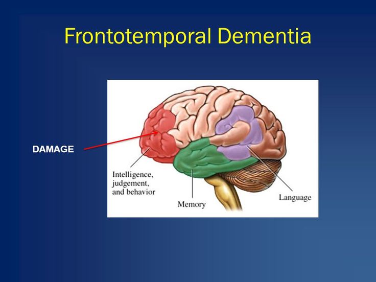
Traditionally, dementia has been viewed as a singular, global decline of mental functions. However it is increasingly clear that this is not the case. Different disease processes affect specific parts of the brain, resulting in diverse patterns of mental decline. The location of the disease determines the symptoms that occur.
As you can see on the diagram, fronto-temporal dementia and semantic dementia affect areas towards the front of the brain, whereas Alzheimers disease affects the areas at the back. The different disease processes are therefore associated with different symptoms.
Patients with Alzheimers disease typically have problems in memory, visual and spatial function, and language. They find it difficult to learn new information, see things around them, and retrieve words in conversation. In contrast to their often debilitating mental decline, they are fully insightful and concerned about their problems, with preserved personality and behaviour.
Patients with fronto-temporal dementia and semantic dementia are entirely different. Since the posterior areas of the brain remain unaffected, there are no problems in visual function. However, the affected frontal areas are responsible for controlling behaviour, meaning that there are often profound changes in personality.
The Course Of Aphasia And Visual Cognitive Impairment

Figure summarizes the course of the disease from its onset. At the age of 74, in addition to fluent spontaneous speech, the frequent use of demonstrative pronouns, as well as roundabout explanations and phonemic paraphasia, were observed. The percentage of correct sentence repetitions was approximately 50%.
Fig. 1
Clinical course of the patient from the onset of the disease. The diagram shows the flow of neurological and neuropsychological processes from the onset of the disease to the appearance of the mirror phenomenon
At the age of 75, nausea appeared and donepezil was discontinued. From around this time, her auditory comprehension declined further and she often asked for repetition. In addition, disinhibited attitudes appeared, such as an uncontrollable urge to reach out and to touch things in front of her, the lack of ability to hold herself quiet in a group, and touching the shoulder or back of the attending physician like a close friend.
Don’t Miss: Can Cause Sores Rashes Dementia Or Blindness
What Gets Stored In A Cookie
This site stores nothing other than an automatically generated session ID in the cookie no other information is captured.
In general, only the information that you provide, or the choices you make while visiting a web site, can be stored in a cookie. For example, the site cannot determine your email name unless you choose to type it. Allowing a website to create a cookie does not give that or any other site access to the rest of your computer, and only the site that created the cookie can read it.
Amyloid Imaging In Dementia With Lewy Bodies
Shimada et al reported that amyloid deposits are associated with AD-like atrophy in patients with DLB/PD with dementia. Patients with DLB have higher amyloid deposits than patients with PD and PD with dementia. Amyloid deposits have been linked to cognitive impairment in DLB, and may contribute to the timing of the onset of dementia relative to that of Parkinsonism in Lewy body dementia. However, as shown in , severe metabolic reduction was seen in some patients with DLB despite no evidence of amyloid deposits. In particular, regional metabolic reduction in patients with DLB was observed in the parietotemporal, posterior cingulate, and frontal association cortices, which are the same regions affected in AD but are not correlated with amyloid deposit.
Also Check: Where To Buy Jelly Drops For Dementia Patients
A Taxonomy Of The Progressive Aphasias
The analysis of spontaneous speech and specific speech and language tasks together allow the patient’s speech syndrome to be defined . While it is usually possible to align the individual case with one of these syndromes predominantly, syndromes commonly overlap and fragmentary syndromes are common. Moreover, each of the syndromes can occur in isolation or as part of a more widespread disorder. PNFA and SD are the most common and the best defined syndromes: they are the canonical subtypes of the progressive aphasias and form part of most clinical classifications of FTLD . Considered as a group, however, the taxonomy of the progressive aphasias remains among the most problematic confronting clinical neurology. Despite these caveats, an appreciation of the relations between the progressive aphasia syndromes and their disease associations helps guide the assessment of the individual patient and the formulation of a differential diagnosis. Here we consider each of the syndromes as they are schematized in .
Studies Of Cognitive Impairment In Older Patients With Epilepsy
There are surprisingly few systematic studies that have addressed the issue of cognitive function specifically in the elderly epilepsy population . Most of these are cross-sectional investigations that tested small samples of patients with young-onset epilepsy , with only one study that has reported on late-onset epilepsy . Nevertheless, the work that has been published indicates that overall older adults with epilepsy have greater deficits compared to healthy older people across cognitive domains, but especially in short and long-term visual and verbal memory, executive functions, attention and psychomotor, or processing speed.
The results revealed that many older individuals with new-onset focal epilepsy were cognitively impaired before initiation of AEDs, with 43% markedly affected, 35% were unimpaired while 6% scored above average. Greater deficits were associated with cerebral infarction or cerebrovascular aetiology, neurological comorbidity and higher body mass index. Subjective performance ratings indicated limited insight into cognitive impairments. These findings underline the importance of early cognitive screening to obtain a baseline assessment allowing quantification of any decline and to evaluate effects of subsequent pharmacological treatment. Overall, the limited existing studies on this topic show that older individuals with epilepsy appear to have significant deficits in cognitive function across the board.
Recommended Reading: Alzheimers Awareness Ribbons
Longitudinal Grey And White Matter Changes In Frontotemporal Dementia And Alzheimers Disease
-
Affiliations Center of Geriatrics and Gerontology, University Medical Center, Freiburg, Germany, Department of Nuclear Medicine, University Medical Center, Freiburg, Germany
-
Affiliation Neuroscience Research Australia, Sydney, Australia
-
Affiliation Neuroscience Research Australia, Sydney, Australia
-
Affiliations Neuroscience Research Australia, Sydney, Australia, School of Medical Sciences, University of New South Wales, Sydney, Australia
-
Affiliation Center of Geriatrics and Gerontology, University Medical Center, Freiburg, Germany
-
Affiliation Swiss Epilepsy Centre, Zürich, Switzerland
-
Affiliations Neuroscience Research Australia, Sydney, Australia, ARC Centre of Excellence in Cognition and its Disorders, Sydney, Australia, School of Medical Sciences, University of New South Wales, Sydney, Australia
-
* E-mail:
Affiliations Neuroscience Research Australia, Sydney, Australia, ARC Centre of Excellence in Cognition and its Disorders, Sydney, Australia, School of Medical Sciences, University of New South Wales, Sydney, Australia
Understanding Parts Of The Brain
Learn about the parts of the brain and how dementia damages them, as well as about the symptoms the damage causes.
Dementia is caused when the brain is damaged by diseases, such as Alzheimers disease or a series of strokes. Alzheimers disease is the most common cause of dementia, but not the only one.
A person with dementia will experience symptoms depending on the parts of the brain that are damaged, and the disease that is causing the dementia.
Also Check: Multi Infarct Dementia Treatment