Enlarged Brain Ventricles Symptoms
Symptoms of hydrocephalus vary with age, disease progression, and individual differences in tolerance to the condition. For example, an infantâs ability to compensate for increased CSF pressure and enlargement of the ventricles differs from an adultâs. The infant skull can expand to accommodate the buildup of CSF because the sutures have not yet closed.
In infancy, the most obvious indication of hydrocephalus is often a rapid increase in head circumference or an unusually large head size. Other symptoms may include vomiting, sleepiness, irritability, downward deviation of the eyes , and seizures.
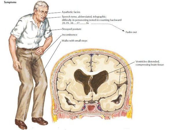
Older children and adults may experience different symptoms because their skulls cannot expand to accommodate the buildup of CSF. Symptoms may include headache followed by vomiting, nausea, blurred or double vision, sun setting of the eyes, problems with balance, poor coordination, gait disturbance, urinary incontinence, slowing or loss of developmental progress, lethargy, drowsiness, irritability, or other changes in personality or cognition including memory loss.
The symptoms described in this section account for the most typical ways in which progressive hydrocephalus is noticeable, but it is important to remember that symptoms vary significantly from person to person.
Regional Gliosis Takes The Place Of Ependyma In Areas Of Lv Expansion
Figure 2. Ventricle expansion is minimal and ependymal cell coverage maintained in cognitively normal individuals . Superior and inferior views of 3D volumetric representations of the LVs for CN1 and CN2 . LV volumes at the indicated ages are overlaid with base volumes shown in blue, expansion in red and stenosis in green. Corresponding immunolabeling of subject-matched biospecimens is shown below 3D representations, with lowercase representing areas of the frontal and occipital horn locations from which tissue was dissected. GFAP highlights regional gliosis in red, AQP4 outlines ependymal cells in blue. Areas of intact ependymal cell coverage are outlined by a white dotted line. Scale bar 50 m.
Figure 3. Ventricle expansion and associated glial scar formation are more rapid and extensive with cognitive impairment. Superior and inferior views of 3D volumetric representations of the LVs for CI1 and CI2 . LV volumes at the indicated ages are overlaid with base volumes shown in blue, expansion in red, and stenosis in green. Corresponding immunolabeling of subject-matched biospecimens is shown below 3D representations, with lowercase representing areas of the frontal and occipital horn locations from which tissue was dissected. GFAP highlights regional gliosis in red, AQP4 outlines ependymal cells in blue. Areas of intact ependymal cell coverage are outlined by a white dotted line. Scale bar 50 m.
What Gives Rise To The Ventricles Of The Brain
What gives rise to the ventricles of the brain? The ventricular system arises from the hollow space within the developing neural tube and gives rise to cisterns within the CNS, from the brain to the spinal cord. In the brain, the ventricular system consists of paired lateral ventricles that connect to the midline third ventricle via bilateral foramina of Monro.
What forms the ventricles of the brain?;The ependymal cell form the lining of the ventricles and are continuous with the epithelium of the choroid plexus. The arachnoid barrier is formed by the outer layer of the cells of the arachnoid, which are joined by tight junctions and have similar permeability to those of the brain blood vessels.
Do brain ventricles grow?;In the brain of a healthy fetus, the ventricles are about 10 millimeters wide. However, CSF may become trapped in the spaces, causing them to grow progressively larger. Ventriculomegaly is a term that describes the actual image of the enlarged spaces as it appears on a prenatal ultrasound.
What do large ventricles in the brain mean?;Hydrocephalus is the abnormal enlargement of the brain cavities caused by a build-up of cerebrospinal fluid . Usually, the body maintains a constant circulation and absorption of CSF. Untreated, hydrocephalus can result in brain damage or death.
Also Check: Does Alzheimer’s Run In Families
What Do Enlarged Brain Ventricles Indicate
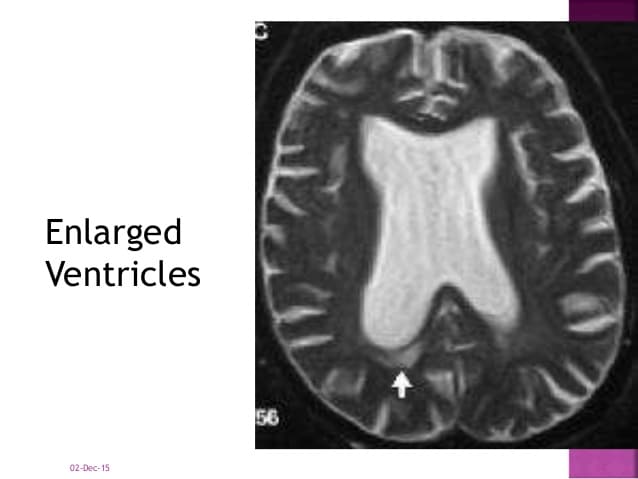
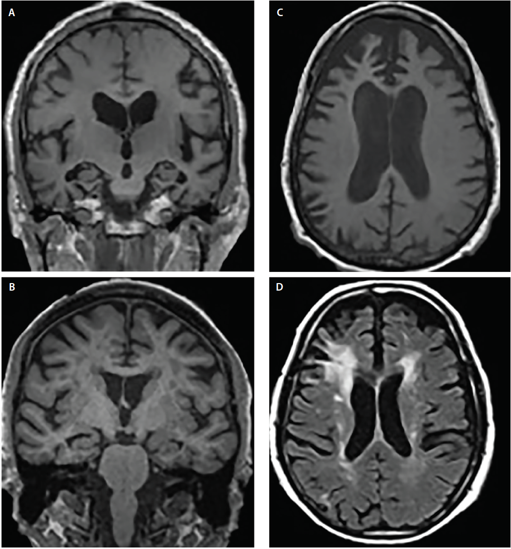
Enlarged ventricles in the brain may be a sign of normal pressure hydrocephalus. It happens when one or more ventricals, which are normally hollow areas in the brain, have too much cerebrospinal fluid.
Cerebrospinal fluid, or CSF, is made and stored in the brain’s ventricles. Its purpose is to help protect the central nervous system and supply it with nutrients. It also removes some toxins and wastes. Any excess CSF should drain away and be absorbed by the body.
If the CSF doesn’t drain, it can build up in the ventricles and cause them to press against the brain. In normal pressure hydrocephalus, this build-up of fluid is gradual. Despite this, there are still symptoms associated with it.
Most people with normal pressure hydrocephalus are over 60. The symptoms can include cognitive changes, clumsiness and incontinence. If the cognitive symptoms are so severe that they disrupt daily life, the patient is said to have dementia. However, normal pressure hydrocephalus, unlike disorders like Parkinson’s disease and Alzheimer’s disease, can be reversed. A shunt can be surgically implanted to drain off the excess CSF.
Sometimes, the condition seems to have no cause or is a complication of a tumor, infection or brain hemorrhage.
Ventricular And Periventricular Anomalies In The Aging And Cognitively Impaired Brain

- 1Department of Physiology and Neurobiology, University of Connecticut, Storrs, CT, United States
- 2Laboratory of Behavioral Neuroscience, Intramural Research Program, National Institute on Aging, Baltimore, MD, United States
- 3Department of Pathology, Johns Hopkins School of Medicine, Baltimore, MD, United States
- 4UConn Center on Aging, University of Connecticut School of Medicine, Farmington, CT, United States
- 5Department of Psychological Sciences, University of Connecticut, Storrs, CT, United States
Also Check: Senile Dementia Of Alzheimer’s Type
What Causes Enlarged Brain Ventricles
The causes of enlarged ventricles of brain are still not well understood. Hydrocephalus may result from inherited genetic abnormalities or developmental disorders . Other possible causes include complications of premature birth such as intraventricular hemorrhage, diseases such as meningitis, tumors, traumatic head injury, or subarachnoid hemorrhage, which block the exit of CSF from the ventricles to the cisterns or eliminate the passageway for CSF within the cisterns.
Support For Normal Pressure Hydrocephalus
Coping with the symptoms of NPH can be difficult for both you and your family members. The condition affects every aspect of your life, including family relationships, work, financial status, social life, and physical and mental health. You may feel overwhelmed, depressed, frustrated, angry, or resentful. These feelings do not help the situation and usually make it worse.
This is why support groups were invented. Support groups are groups of people who are going through the same things and want to help themselves and others by sharing coping strategies.
Support groups meet in person, on the telephone, or on the internet. To find a support group that works for you, contact the organizations listed below. You can also ask your health care provider or behavior therapist, or go on the internet. If you do not have access to the internet, go to the public library.
For more information about support groups, contact the following agencies:
- Family Caregiver Alliance, National Center on Caregiving — 445-8106
- Hydrocephalus Association — 732-7040 or 598-3789
- National Hydrocephalus Foundation — 924-6666
- Hydrocephalus Support Group, Inc. — 532-8228
Recommended Reading: Moving A Parent To Memory Care
How Many Ventricles Are In The Brain
Four. The right and left lateral ventricles, third ventricle and the fourth ventricle. The ventricular system is made up of four ventricles connected by narrow passages. Normally, cerebrospinal fluid flows through the ventricles, exits into cisterns at the base of the brain, bathes the surfaces of the brain and spinal cord, and then reabsorbs into the bloodstream.
What Is The Difference Between Right And Left Ventricle
The right ventricle passes the blood on to the pulmonary artery, which sends it to the lungs to pick up oxygen. The left atrium receives the now oxygen-rich blood from the lungs and pumps it into the left ventricle. The left ventricle pumps the oxygen-rich blood to the body through a large network of arteries.
Also Check: Sandyside Senior Living
Ventricular Tracing And Extraction
Ventricular extraction followed a semi-automated ventricular segmentation approach. Briefly, an experienced human rater traced the lateral ventricles of four subjects in three partitions per hemisphere â superior horn, temporal horn and ventricular body/occipital horn. These traces were converted into 3D parametric ventricular mesh models, termed atlases. Next each of the four atlases was fluidly registered to each unsegmented study image. The resulting four ventricular segmentations per study subject were averaged resulting in one final 3D ventricular model for each study participant. Averaging four automated segmentations for each subject minimizes as much as possible automated labeling errors and most accurately captures individual anatomy.
Imaging Data Acquisition And Preprocessing
Imaging data were collected on a 1.5 T Signa MRI scanner with the following protocol: 3D spoiled gradient echo, gapless coronal acquisition, TR 28 msec, TE 6 msec, FOV 220 mm, 256 Ã192 matrix, slice thickness 1.5 mm, voxel size 0.9Ã0.9Ã1.5 mm . We used the Minctracc algorithm to rotate and globally scale all MRI scans to match the International Consortium for Brain mapping ICBM53 average brain imaging template using a 9-parameter linear transformation and a regularized tricubic B-spline approach for image intensity inhomogeneity correction. The post-processed images had a reconstructed isotropic voxel dimension of 1Ã1Ã1 mm. The ICBM53 was chosen over the ICBM152 and the ICBM305 templates as it has better contrast and more sharply defined cortical and subcortical structural definition, which may be helpful for improved cross-subject image registration.
Also Check: Senility Vs Dementia Vs Alzheimer’s
What Are The Symptoms
The symptoms of hydrocephalus can vary significantly from person to person and mostly depend on age. ;Conditions other than hydrocephalus can cause similar symptoms so it is important to see a doctor to receive proper diagnosis and treatment.InfantsSigns and symptoms of hydrocephalus in infants include:
- a rapid increase in head size
- an unusually large head
Brain imaging and other testsTests to accurately diagnose hydrocephalus and rule out other conditions may include:
What Separates The Lateral Ventricles Of The Brain

4.3/5lateral ventriclesthe answer
Lateral VentricleThe 2 lateral ventricles are each other by a thin vertical sheet of nervous tissue called septum pellucidum covered on either side by ependyma. It communicates with the third ventricle through the interventricular foramen of Monro.
Furthermore, where are the lateral ventricles of the brain located? The left and right lateral ventricles are located within their respective hemispheres of the cerebrum. They have ‘horns’ which project into the frontal, occipital and temporal lobes. The volume of the lateral ventricles increases with age.
Additionally, what are the lateral ventricles in the brain?
The right and left lateral ventricles are structures within the brain that contain cerebrospinal fluid, a clear, watery fluid that provides cushioning for the brain while also helping to circulate nutrients and remove waste.
What are the four ventricles of the brain and their functions?
The ventricles of the brain are a communicating network of cavities filled with cerebrospinal fluid and located within the brain parenchyma. The ventricular system is composed of 2 lateral ventricles, the third ventricle, the cerebral aqueduct, and the fourth ventricle .
Recommended Reading: Difference Between Senility And Dementia
What Is The Purpose Of Ventricles
The right ventricle pumps the oxygen-poor blood to the lungs through the pulmonary valve. The left atrium receives oxygen-rich blood from the lungs and pumps it to the left ventricle through the mitral valve. The left ventricle pumps the oxygen-rich blood through the aortic valve out to the rest of the body.
Enlarged Brain Ventricles Treatment
Hydrocephalus is most often treated by surgically inserting a shunt system. This system diverts the flow of CSF from the CNS to another area of the body where it can be absorbed as part of the normal circulatory process.
A shunt is a flexible but sturdy plastic tube. A shunt system consists of the shunt, a catheter, and a valve. One end of the catheter is placed within a ventricle inside the brain or in the CSF outside the spinal cord. The other end of the catheter is commonly placed within the abdominal cavity, but may also be placed at other sites in the body such as a chamber of the heart or areas around the lung where the CSF can drain and be absorbed. A valve located along the catheter maintains one-way flow and regulates the rate of CSF flow.
A limited number of individuals can be treated with an alternative procedure called third ventriculostomy. In this procedure, a neuroendoscope â a small camera that uses fiber optic technology to visualize small and difficult to reach surgical areas â allows a doctor to view the ventricular surface. Once the scope is guided into position, a small tool makes a tiny hole in the floor of the third ventricle, which allows the CSF to bypass the obstruction and flow toward the site of resorption around the surface of the brain.
What are the possible complications of a shunt system?
Also Check: What Is The Difference Between Dementia And Senility
How Does The Brain Change Over The Course Of Alzheimer’s
by CNRS
What changes in the brain are caused by Alzheimer’s disease? How do these changes differ from those observed in the normal ageing process? Researchers from the CNRS, the École pratique des hautes études and the University of Valencia explored these questions by analysing over 4,000 MRI scans of healthy and diseased brains using the “volBrain” platform. Their models, published in the March 8, 2019 edition of Scientific Reports, reveal an early atrophy of the amygdala and hippocampus at age 40 in patients with Alzheimer’s disease.
Alzheimer’s disease is characterised by changes in the brain, some of which can be measured in the patient using biomarkers such as the size of different areas of the brain. Studying these biomarkers has shown that certain brain structures shrink in the presence of Alzheimer’s well before the first signs of dementia appear. Key questions remained however: when and how did changes in these biomarkers differ between a healthy subject and one with Alzheimer’s, for example?
Developed by the CNRS, the Université de Bordeaux and the University of Valencia in Spain, this free platform allows researchers around the world to upload structural MRI files and obtain in record time an automatic analysis of the scanned brain structure volumes. Since its creation in 2015, over 110,000 brain MRIs have been analysed for over 2,500 users worldwide.
Explore further
Where Can I Get More Information
For more information on neurological disorders or research programs funded by the National Institute of Neurological Disorders and Stroke, contact the Institute’s Brain Resources and Information Network at:
Office of Communications and Public LiaisonNational Institute of Neurological Disorders and StrokeNational Institutes of HealthBethesda, MD 20892
NINDS health-related material is provided for information purposes only and does not necessarily represent endorsement by or an official position of the National Institute of Neurological Disorders and Stroke or any other Federal agency. Advice on the treatment or care of an individual patient should be obtained through consultation with a physician who has examined that patient or is familiar with that patient’s medical history.
All NINDS-prepared information is in the public domain and may be freely copied. Credit to the NINDS or the NIH is appreciated.
You May Like: Senile Dementia Of The Alzheimer Type
What Is The Best Treatment For Hydrocephalus
The most common treatment for hydrocephalus is the surgical insertion of a drainage system, called a shunt. It consists of a long, flexible tube with a valve that keeps fluid from the brain flowing in the right direction and at the proper rate. One end of the tubing is usually placed in one of the brains ventricles.
Can Ventriculomegaly Be Treated
How is ventriculomegaly treated? Ventriculomegaly only needs to be treated if the baby has hydrocephalus, a build-up of cerebrospinal fluid in the brain. Treatments for hydrocephalus include: Shunt placement: In this process a small tube is implanted in the brain while the child is under anesthesia.
You May Like: How Does Dementia Kill You
Hydrofracking Ventriculomegaly And Brain Atrophy
Researchers suspect that enlarged ventricles, known as ventriculomegaly,; seen in many neurodegenerative diseases may be the result of atrophy of the brain. The cause of the damage or atrophy of the brain may be;due to destructive waves and hydraulic pressures that damage tissues by a process I compare to;hydrofracking which is used by engineers to fracture rocks. Ventriculomegaly and brain atrophy have been associated with Alzheimers disease, Parkinsons disease and its variants, called Parkinsons Plus, as well as multiple sclerosis, amyotrophic lateral sclerosis and Huntingtons disease.
The picture above shows the left half of the brain. The face would be to the right. The cauliflower structure in the lower left corner is the cerebellum. The hollow area in the middle of the brain is the left lateral ventricle. The heavy white structure that forms the roof over the lateral ventricle;is;the corpus callosum. The heavy white structure that forms the floor is;the fornix. The corpus callosum is a group of;myelinated ;high speed interconnecting communication pathways that link the left and right halves of the brain. The fornix is, likewise, a high-speed communication pathway of white fibers. The third ventricle is located just;below the fornix. The fourth ventricle is the space shaped like a dart between the cerebellum and brainstem. The ventricles are chambers in the core of the brain and brainstem where cerebrospinal fluid ;is produced.
What Are The Different Types Of Hydrocephalus

Hydrocephalus can affect anyone at any age but is most common in infants and older adults. ;Some of these cases can be associated with abnormalities in the brain and spinal cord during pregnancy. ;The two major types of hydrocephalus are called communicating hydrocephalus and non-communicating hydrocephalus.
Communicating hydrocephalus occurs when the flow of CSF is blocked after it exits the ventricles. This form is called communicating because the CSF can still flow between the ventricles, the passages between which remain open.; Reduced flow and absorption of CSF into specialized blood vessels called arachnoid villi can also result in a buildup of CSF in the ventricles and communicating hydrocephalus.;
- Non-communicating hydrocephalus happens when the flow of CSF is blocked along one or more of the narrow passages connecting the ventricles. ;
Two additional types of hydrocephalus include:
Also Check: Does Medicare Cover Nursing Home Care For Dementia