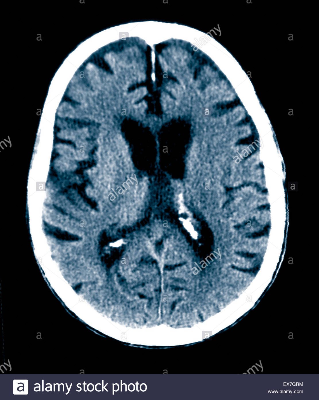
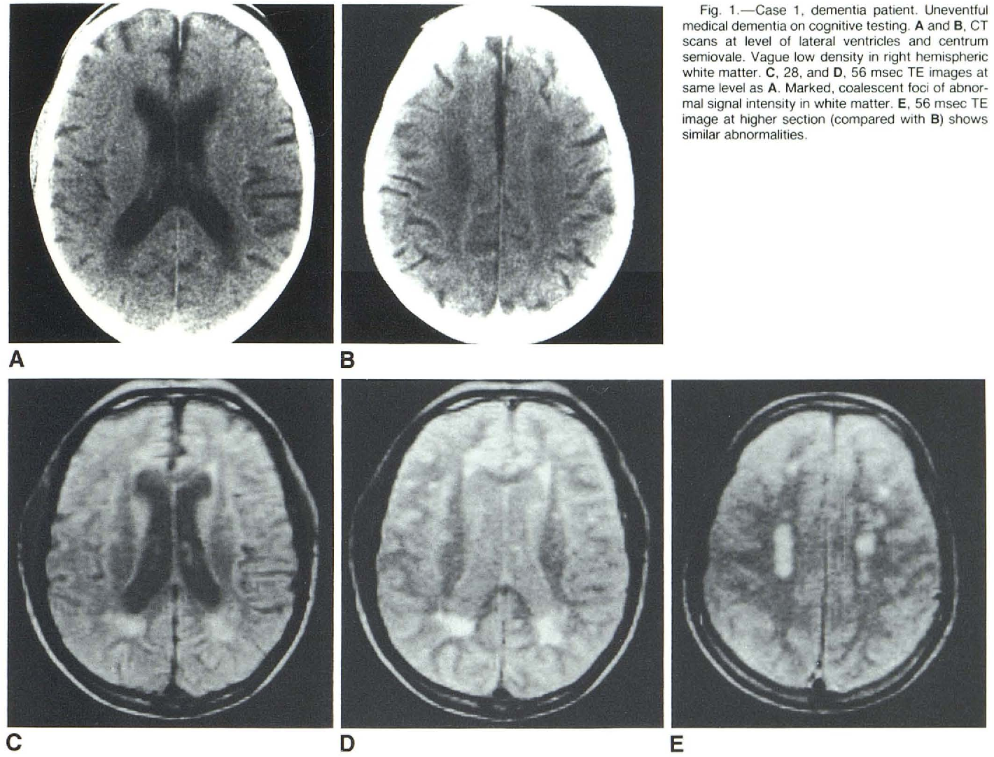
Mri Scans Can Show Dementia
According to researchers from Perelman School of Medicine at the University of Pennsylvania, the answer to can an MRI detect dementia is to some extent yes.
The scientists explained that doctors have an easier time telling whether a person has dementia through MRI scans.
This gets rid of the need to carry out invasive tests that people find unfriendly like the lumbar puncture where a doctor must stick a needle in the spine.
Additionally, it also helps to speed up the diagnosis process which is important seeing that dementia diagnosis for the longest time has been a struggle for medics often leading to delayed treatment.
In addition to telling whether a person has dementia, MRI scans may in the future help doctors determine whether an individual is at risk of dementia according to new research.
Research from the University of California San Francisco and the Washington University School of Medicine in St. Louis conducted a small study where MRI brain scans were able to predict with 89% accuracy the people who were going to develop dementia in three years.
The researchers presented their findings in Chicago during a Radiological Society of North America meeting.
It suggested that in a few years, physicians will be able to tell people their risk of developing dementia before they start to showcase any symptoms of the neurodegenerative illness.
What Is The Main Cause Of Alzheimers Disease
We wont be wrong to say that the Alzheimers disease comes with age, which is why it is the main cause. However, just like all types of dementia, the actual cause related to Alzheimers is the death of brain cells.
Alzheimers disease is a neurodegenerative one, which means that there is progressive brain cell death which happens over time. In a nutshell, the brain tissue of the person suffering from it starts losing its nerve cells and connections, resulting in this disease . Read more about what causes Alzheimers.
Devoted Guardians’ Response to COVID-19
Devoted Guardians is actively monitoring the progression of the coronavirus, COVID-19, to ensure that we have the most accurate and latest information on the threat of the virus. As you know, this situation continues to develop rapidly as new cases are identified in our communities and our protocols will be adjusted as needed.
While most cases of COVID-19 are mild, causing only fever and cough, a very small percentage of cases become severe and may progress particularly in the elderly and people with underlying medical conditions. Because this is the primary population that Devoted Guardians serves, we understand your concerns and want to share with you how our organization is responding to the threat of COVID-19.
Measure Volume In The Brain
An MRI can provide the ability to view the brain with 3D imaging. It can measure the size and amount of cells in the hippocampus, an area of the brain that typically shows atrophy during the course of Alzheimer’s disease. The hippocampus is responsible for accessing memory which is often one of the first functions to noticeably decline in Alzheimer’s.;
An MRI of someone with Alzheimer’s disease may also show parietal atrophy. The parietal lobe of the brain is located in the upper back portion of the brain and is responsible for several different functions including visual perception, ordering and calculation, and the sense of our body’s location.
You May Like: Does Alzheimer Disease Run In The Family
Utility Of Functional Mri In The Study Of Ad
A relatively small number of fMRI studies have been published in subjects at risk for AD, including MCI subjects and genetic at-risk individuals yielding somewhat discrepant findings. Several studies have reported decreased mesial temporal lobe activation in MCI and genetic at-risk subjects . Interestingly, several fMRI studies have reported evidence of increased MTL activity in at-risk subjects, particularly among very mild MCI subjects , and cognitively intact individuals with genetic risk for AD . It is likely that these discrepant results are related to specific paradigm demands, stage of impairment, and behavioral performance. A common feature of the studies reporting evidence of increased fMRI activity is that the at-risk subjects were able to perform the fMRI tasks reasonably well. In particular, the event-related fMRI studies have found that hyperactivity was observed specifically during successful memory trials, which suggested that hyperactivity might represent a compensatory mechanism in the setting of early AD pathology .
Group map of fMRI activity showing regions that increase activity or decrease activity during successful encoding. Group map of 11C-PiB retention in a group of non-demented older individuals. Note the anatomic overlap of PiB retention to default network .
Pet Imaging May Also Be Used While Diagnosing Alzheimer’s

Positron-Emissions Tomography, or the PET scan, is the last type of investigation that we will spotlight in this article. There is no need to get into the complicated physics of what is happening to explain how this instrument works, but the simple version may be helpful. A PET scan;is a type of study that is used to determine how much glucose is being used by tissue. This can give a reasonable representation of how quickly the brain is metabolizing the sugar. The more active an area, the more glucose will be needed in order to maintain these functions.;
The application of this PET scan in the diagnosis of Alzheimer’s Disease is apparent in modern medicine. There are numerous studies that have shown the PET scan to be a very good tool in diagnosing the early changes in the brain. It is also able to differentiate more precisely between normal aging and patterns likely seen only in patients who have Alzheimer’s Disease, making it a fairly accurate diagnostic tool. Another advantage of this type of investigation is that it can produce models that can predict the speed of memory decline in the future. This can help doctors target specific treatments and identify symptoms that may become more pronounced depending on the region of the brain involved in the patient.;
Don’t Miss: Does Medicare Cover Nursing Home Care For Dementia
Imaging In The Diagnosis And Prognosis Of Ad
The uncertainty inherent in a clinical diagnosis of AD has driven a search for diagnostic imaging markers. A definitive diagnosis still requires histopathological confirmation and the inaccessibility of the brain means imaging has a key role as a window on the brain. Historically, imagingfirst computed tomography and then MRIwas used only to exclude potentially surgically treatable causes of cognitive decline. Now its position in diagnosis also includes providing positive support for a clinical diagnosis of AD in symptomatic individuals by identifying characteristic patterns of structural and functional cerebral alterations. We can now also visualize the specific molecular pathology of the diseaseamyloid depositswith amyloid imaging. Alongside this increasing specificity for AD, imaging also contributes to differential diagnosis in practice by identifying alternative and/or contributory pathologies. Imaging is central to identifying vascular and non-AD degenerative pathologies and has helped in the recognition of the prevalence of mixed pathology in dementia.
The Changing Roles And Scope Of Neuroimaging In Alzheimer Disease
There has been a transformation in the part played by neuroimaging in Alzheimer disease research and practice in the last decades. Diagnostically, imaging has moved from a minor exclusionary role to a central position. In research, imaging is helping address many of the scientific questions outlined in : providing insights into the effects of AD and its temporal and spatial evolution. Furthermore, imaging is an established tool in drug discovery, increasingly required in therapeutic trials as part of inclusion criteria, as a safety marker, and as an outcome measure.
Concomitantly the potential of brain imaging has expanded rapidly with new modalities and novel ways of acquiring images and of analysing them. This article cannot be comprehensive. Instead, it addresses broad categories of structural, functional, and molecular imaging in AD. The specific modalities included are magnetic resonance imaging and positron emission tomography . These modalities have different strengths and limitations and as a result have different and often complementary roles and scope.
Also Check: How Does Dementia Kill You
How Brain Scans Assist With Identifying Dementia
Alzheimers disease is the most common type of dementia. A CT scan or MRI can assist with identifying a physical change or brain condition contributing to dementia or Alzheimers symptoms: These signs include:
- Indication a stroke has occurred
- Tumors
- Cortical atrophy, or wrinkled ridges of tissue forming on the brain
- Changes in the brains structure and functioning, including loss of brain mass
While dementia may be a symptom of the above conditions, an atrophied hippocampus is a sign of Alzheimers disease.
While preferred, CT and MRI scans might not deliver the results a doctor is seeking. If tests come back inconclusive, positron emission tomography and single-photon emission computed tomography might be requested. These scans examine various aspects of brain activity, including oxygen use and blood flow, and can identify symptoms that separate Alzheimers from other types of dementia.
Additionally, an electroencephalogram may be requested to observe abnormal brain activity. While an EEG is not ideal for diagnosing Alzheimers disease, it can identify other conditions for which dementia and cognitive decline are symptoms. Furthermore, this test can detect the source of seizures an issue for roughly 10 percent of Alzheimers patients.
Who Diagnoses Dementia
The General Practitioner is usually the first contact when concerns about thinking or memory arise. The GP will take a medical history and may carry out a brief test of memory and concentration. If the GP is concerned about the possibility of dementia, the person may be referred to a specialist or specialist memory centre. It is important to remember that the choice of doctor is up to you so if after your visit you are still concerned and wish a referral to a specialist, you may wish to ask for a second opinion.;;
Specialists such as neurologists, geriatricians, psychogeriatricians, psychiatrists, and neuropsychologists have a more detailed knowledge of the memory and behaviour changes associated with dementia and may perform or arrange in-depth assessments, brain scans and blood tests. In Australia, a specialist must confirm the diagnosis of Alzheimers disease in order for you to be eligible for subsidised Alzheimers medications.;;
Aged Care Assessment Teams are multidisciplinary teams often comprised of social workers, occupational;therapists, as well as nurses and doctors. ACATs are usually based in hospitals or regional community health centres. ACATs assess the health needs of ageing individuals, put the individual in contact with relevant services, make recommendations about the level of care required and approve eligibility for certain services.;;
You May Like: What Is The Difference Between Dementia And Senility
How To Refer A Patient
Please contact us so that we can learn more about your needs.
For those who need a referral for a scan through an Alzheimer’s specialist, please contact the UCSF Memory and Aging CenterPh: ; 353-2057Fax: 353-8292
Neurologists, gerontologists and psychiatrists participating in the IDEAS* study ;can refer directly to UCSF Imaging;by calling Radiology Scheduling at; 353-3900. All others, please contact the UCSF Memory and Aging Center at 353-2057.
How Brain Scans Help Diagnose Alzheimers Disease
- 8.31.20
- Dementia Care
When the signs are recognized, Alzheimers disease can be identified in the early stages. However, even when forgetfulness and other indicators are present, the diagnostic process is not always straightforward.
Alzheimers disease cannot be determined by a single blood test and the evaluation often requires multiple specialists to consult.
Also, one set of tests may not be enough. An evaluation may need to take place within six months to a year to observe the diseases potential progression.
Read Also: How To Move A Parent With Dementia To Assisted Living
Why Early Detection Can Be Difficult
Alzheimers disease usually is not diagnosed in the early stages, even in people who visit their primary care doctors with memory complaints.
- People and their families generally underreport the symptoms.
- They may confuse them with normal signs of aging.
- The symptoms may emerge so gradually that the person affected doesnt recognize them.
- The person may be aware of some symptoms but go to great lengths to conceal them.
Recognizing symptoms early is crucial because medication to control symptoms is most effective in the early stages of the disease and early diagnosis allows the individual and his or her family members to plan for the future. If you or a loved one is experiencing any of the following symptoms, contact a physician.
Early Warning Signs And Diagnosis

Alzheimers Disease can be caught in the early stageswhen the best treatments are availableby watching for telltale warning signs. If you recognize the warning signs in yourself or a loved one, make an appointment to see your physician right away. Brain imaging technology can diagnose Alzheimers early, improving the opportunities for symptom management.
Don’t Miss: Senile Dementia Of The Alzheimer Type
Proposed Treatments Of Patients Having Ad With Ionizing Radiation
Bistolfi states that vascularcerebral amyloidosis is the hallmark of AD. Localized tracheobronchial amyloidosis has been successfully treated with beams of radiation, 20 Gy in 10 fractions of 200 cGy in 2 weeks. As 20 Gy in 2 weeks is followed by inflammatory reactions, this high dosage cannot be suggested in the hypothetical treatment of AD. An innovative alternative might be a weekly long-term low dose, say 50 to 100 cGy, fractionated radiotherapy , matching the very slow response of amyloid to radiation. Before applying it to patients with AD, the proposed schedule should be tried in patients with TBA to compare the new results of long-term fractionated RT with the results of 20 Gy/2 w. Should long-term fractionated RT prove effective, its application to patients with AD might become an effective and safe treatment.
On July 17, 2013, an application for a patent was published, titled Radiation therapy for treating Alzheimers disease. It makes 14 claims for treating human patients by a method, which is based on studies carried out using mice. The method comprises administering a relatively large amount of ionizing radiation to the brain of the patient employing a variety of different radiation sources. The total dose ranges from 300 to 1800 cGy, administered in dose fractions of 50 to 300 cGy per day. The method is claimed to treat AD by reducing the number or size of amyloid plaques in the brain of the patient.
Current Practice In Diagnosing Dementia
The remainder of this information will provide an overview of the diagnosis process and a guide to what happens after diagnosis.;
It is important to remember that there is no definitive test for diagnosing Alzheimers disease or any of the other common causes of dementia. Findings from a variety of sources and tests must be pooled before a diagnosis can be made, and the process can be complex and time consuming. Even then, uncertainty may still remain, and the diagnosis is often conveyed as possible or probable. Despite this uncertainty, a diagnosis is accurate around 90% of the time.;
People with significant memory loss without other symptoms of dementia, such as behaviour or personality changes, may be classified as having a Mild Cognitive Impairment . MCI is a relatively new concept and more research is needed to understand the relation between MCI and later development of dementia. However, MCI does not necessarily lead to dementia and regular monitoring of memory and thinking skills is recommended in individuals with this diagnosis.;;
Also Check: Senility Vs Dementia Vs Alzheimer’s
Physical And Neurological Examination
Your doctor will check your vital signs, like heart rate, blood pressure, pulse rate, and temperature. A neurological exam can include tests to check your reflexes, balance, coordination, speech, sight, and hearing.
Your doctor may also do a series of tests to evaluate your cognitive abilities and brain function. These tests may feel like a series of puzzles or games that are designed to test your:
- Memory
- Communication and language skills
- Reasoning and planning abilities
These tests can help your doctor determine whether its safe for you to live independently, manage your finances, or drive a vehicle.
If you are diagnosed with Alzheimers, you may need to do these tests periodically to evaluate the condition’s progression.;
Over the course of your appointment, your doctor will also evaluate your behavior as well as your mental and emotional state. On subsequent visits, they will make a note of any changes in your personality or behavior.
Additional Tests To Treat And Manage Dementia
Once an individual is diagnosed with dementia, the next step that follows is helping them understand how the condition will affect them and how to manage it.
Several other medical assessments exist to help physicians understand how the condition affects a specific person. And also help families, as well as caregivers, figure out the best course of treatment for the individual.
Did you HEAR of the peanut butter test?
Neuropsychological Testing
Neuropsychologists and psychologists who have specialized training can also prescribe neuropsychological tests to detect dementia.
The process involves WRITTEN and ORAL tests that can take several hours to complete.
They use these methods to assess the cognitive functions of the person suspected to have dementia.
It helps them figure out if certain areas are impaired.
The tests assess aspects like vision-motor, memory, comprehension, reasoning, coordination, and writing abilities.
Physicians may administer additional tests to find out if the person in question is SUFFERING from mood problems or dementia.
Functional Assessments
Dementia is a cognitive disorder that affects the afflicted persons daily functioning in different regards.
Objective assessments can establish what a person is STILL ABLE to do versus what they can no longer do in light of the condition.
Family members are asked to fillquestionnaires that provide details about the persons daily life in terms of the activities they are able to perform.
Psychosocial evaluation
Recommended Reading: Sandyside Senior Living
Amyloid Pet Scan For Alzheimer’s Disease Assessment
Diagnosing Alzheimers is complex. With no single test currently available, diagnosis is based on an individuals history, physical examination, and cognitive testing. Amyloid PET imaging represents a potential major advance in the assessment of those with cognitive impairment. The scan visualizes plaques present in the brain, which are prime suspects in damaging and killing nerve cells in Alzheimer’s. Before amyloid PET, these plaques could only be detected by examining the brain at autopsy.;
UCSF Imaging’s scientists and physicians are recognized world leaders in the;translation of Alzheimer’s research to clinical care.;Since 2003, UCSF Imaging has offered Alzheimers disease assessment with amyloid PET scanning for patients with memory complaints.;Amyloid PET scanning makes amyloid plaques “light up” on a brain PET scan, enabling, for the first time, accurate detection of plaques in living people. The Department and the multidisciplinary initiatives that serve Alzheimer’s patients, including the UCSF Memory and Aging Center, the Center for Imaging of Neurodegenerative Diseases , and the Alzheimer’s Disease Neuroimaging Initiative , participate;in multiple national research studies;including the IDEAS ;Study. UCSF is one of the worlds largest centers for evaluation of cognitive decline and dementia. We look forward to learning more about your interest in this topic and welcome you to;contact;us any time.