How Mri Is Used To Detect Alzheimer’s Disease
One way to test for Alzheimer’s disease is to assess the brain’s functioning. There are several frequently used cognitive screenings that can be used to evaluate someone’s memory, executive functioning, communication skills, and general cognitive functioning. These tests are commonly done in your healthcare provider’s office widely used is Mini Mental Status Exam or Montreal Cognitive Assessment . These can be very helpful in identifying if a problem exists, or if there’s just a normal lapse in memory.
These can be very helpful in identifying if a problem exists, or if there’s just a normal lapse in memory due to aging. There are, however, several different types of dementia, as well as other conditions that can cause symptoms of dementia but are reversible. There are ways you can tell.
Tracking Progression In Clinical Trials
The search for a valid marker to track disease progression should be viewed in the context of the development of drugs with potential disease-modifying effects. A valid marker of disease activity that has higher measurement precision than the currently used outcomes might provide a surrogate outcome measure. For a biomarker to be accepted as a surrogate outcome in a clinical drug trial, it must both be correlated with the clinical outcome and fully capture the net effect of the intervention on the clinical efficacy outcome., Imaging outcomes could potentially allow meaningfully powered phase II and III clinical trials with significantly smaller patient groups and/or shorter follow-up times than are currently possible, thereby providing a boost to the efficacy of drug development programs. No widely accepted, valid surrogate outcome of disease progression currently exists, although preliminary data indicate that imaging measures could provide adequate power to clinical trials with far smaller samples of patients than are required if traditional cognitive and functional measures are used.,,
Brain Imaging Tests Used To Diagnose Dementia
Although a brain scan alone does not diagnose dementia, it can be a strong indicator when included as part of a wider assessment. Once simpler tests, such as blood tests and mental ability tests, have been used to eliminate other possible conditions, a brain scan can confirm dementia as the patients diagnosis.
In addition to helping diagnose dementia, a brain imaging test can check for other potential causes of common dementia symptoms, such as a brain tumor or stroke. If the symptoms are coming from a different source, they will require a different treatment, so an accurate diagnosis is imperative. With imaging tests, patients can be more confident they are receiving the correct treatment and following the right recovery plan.
While there is a variety of brain imaging tests patients could undergo, below are the most common for diagnosing possible cases of dementia.
Recommended Reading: Aphasia In Alzheimer’s
Early Warning Signs And Diagnosis
Alzheimers Disease can be caught in the early stageswhen the best treatments are availableby watching for telltale warning signs. If you recognize the warning signs in yourself or a loved one, make an appointment to see your physician right away. Brain imaging technology can diagnose Alzheimers early, improving the opportunities for symptom management.
Referral To A Specialist

If a GP is unsure about whether you have Alzheimer’s disease, they may refer you to a specialist, such as:
- a psychiatrist
- an elderly care physician
- a neurologist
The specialist may be based in a memory clinic alongside other professionals who are experts in diagnosing, caring for and advising people with dementia and their families.
There’s no simple and reliable test for diagnosing Alzheimer’s disease, but the staff at the memory clinic will listen to the concerns of both you and your family about your memory or thinking.
They’ll assess your memory and other areas of mental ability and, if necessary, arrange more tests to rule out other conditions.
Read Also: Ribbon Color For Dementia
What Is Alzheimers Disease
Alzheimers is thought to be the result of beta-amyloid plaques, which are thick protein deposits present in the brain, and neurofibrillary tangles abnormal structures, which form in neurons building up in the brain. This buildup causes neurons in the brain to cease working, losing connection with other neurons before dying. However, the exact cause of Alzheimers is still unknown. The condition is the most common form of dementia and can quickly progress from being mild to severe.
There are no known causes for Alzheimers since the disease is still being studied. A persons chances of developing Alzheimers tend to increase with age, but people in their 40s and 50s can begin showing symptoms of early-onset Alzheimers. People who have a relative with the condition may also be at higher risk of developing it themselves since the disease has hereditary factors.
A persons overall physical health may also be a factor in whether they develop Alzheimers disease. One study showed that people who have heart disease or other cardiovascular issues could be at a higher risk of developing Alzheimers than those with good heart health. This is because cardiovascular issues often reduce the amount of blood the brain receives, which may increase the cognitive issues associated with Alzheimers.
How Accurate Is Magnetic Resonance Imaging For The Early Diagnosis Of Dementia Due To Alzheimer’s Disease In People With Mild Cognitive Impairment
Why is improving Alzheimer‘s disease diagnosis important?
Cognitive impairment is when people have problems remembering, learning, concentrating and making decisions. People with mild cognitive impairment generally have more memory problems than other people of their age, but these problems are not severe enough to be classified as dementia. Studies have shown that people with MCI and loss of memory are more likely to develop Alzheimer’s disease dementia than people without MCI . Currently, the only reliable way of diagnosing Alzheimer’s disease dementia is to follow people with MCI and assess cognitive changes over the years. Magnetic resonance imaging may detect changes in the brain structures that indicate the beginning of Alzheimer’s disease. Early diagnosis of MCI due to Alzheimer’s disease is important because people with MCI could benefit from early treatment to prevent or delay cognitive decline.
What was the aim of this review?
To assess the diagnostic accuracy of MRI for the early diagnosis of dementia due to Alzheimer’s disease in people with MCI.
What was studied in the review?
The volume of several brain regions was measured with MRI. Most studies measured the volume of the hippocampus, a region of the brain that is associated primarily with memory.
What are the main results in this review?
How reliable are the results of the studies?
Who do the results of this review apply to?
What are the implications of this review?
How up to date is this review?
Don’t Miss: Alzheimers Ribbon Color
Basics Of Amyloid Pet As It Is Applied To Ad
With regard to specific amyloid imaging agents, this review will discuss amyloid tracers in general, while acknowledging that most of the statements are derived from data on the most widely evaluated PET tracer, PiB . At the time of writing, there have been one or two, small published studies using each of the fluorine-18-labelled tracers, florbetaben , florbetapir and flutemetamol in AD patients. Although the PiB PET findings may ultimately be found to extend to these F-18-labeled tracers as well, this cannot be assumed until appropriate studies have been repeated with each individual tracer or until pharmacological equivalency to PiB has been established by direct comparison in the same subjects.
Neuroimaging: Visualising The Brain
Neuroimaging describes a range of tools which are used to visualise the living brain, including computerised tomography scans, magnetic resonance imaging , single photon emission computerised tomography and positron emission tomography .
Researchers are working on new ways of using neuroimaging tools to diagnose Alzheimer’s disease and other types of dementia.
Positron Emission Tomography
In 2004, researchers successfully viewed beta-amyloid plaque deposits in the living human brain. The study used Pittsburgh Compound-B , a substance which binds to amyloid and can be visualised with PET scanning. The results demonstrated that people with Alzheimer’s disease displayed more amyloid deposits in certain brain areas compared to people without the condition. More recent research has shown that PiB-PET can also detect the early brain changes of Alzheimer’s disease before symptoms become apparent.
While PiB has proved quite effective, its widespread clinical use may be limited by the need for specialised equipment to produce PiB at the site of the PET scanner. Researchers are currently developing and testing other compounds that bind to beta-amyloid and may overcome the limitations of PiB.
Magnetic Resonance Imaging
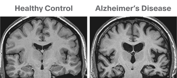
MRI is able to image the structure of the brain, which changes in dementia, to a very high resolution.
Recommended Reading: Does Diet Coke Cause Alzheimer’s
Future Directions In Diagnosis Research
Considerable research effort is being put into the development of better tools for accurate and early diagnosis. Research continues to provide new insights that in the future may promote early detection and improved diagnosis of dementia, including:
- Better dementia assessment tests that are suitable for people from diverse educational, social, linguistic and cultural backgrounds.
- New computerised cognitive assessment tests which can improve the delivery of the test and simplify responses.
- Improved screening tools to allow dementia to be more effectively identified and diagnosed by GPs.
- The development of blood and spinal fluid tests to measure Alzheimers related protein levels and determine the risk of Alzheimers disease.
- The use of sophisticated brain imaging techniques and newly developed dyes to directly view abnormal Alzheimers protein deposits in the brain, yielding specific tests for Alzheimers disease.
How Are Mris Used In The Diagnosis Of Alzheimers Disease
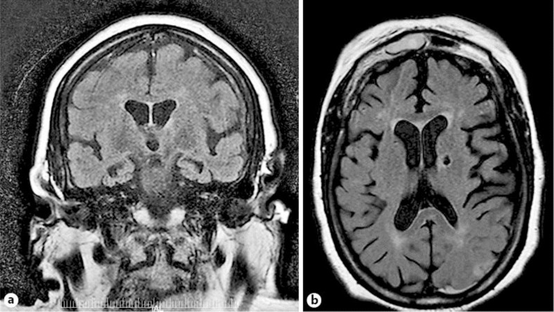
The 3D imaging that MRIs for Alzheimers provide makes it easy for physicians to spot abnormalities in the brain. These abnormalities can be potential markers of Alzheimers, but they could also be symptoms of other cognitive conditions some of which may even be reversible if caught in time.
One of the first areas of the brain to be affected by Alzheimers disease is the hippocampus, which is responsible for developing new memories and helps your brain store them. When the hippocampus shrinks as a result of Alzheimers, the brains ability to form and recall memories diminishes. An Alzheimers MRI produces 3D imaging of the hippocampus, clearly showing how many cells are present and how big it is.
The parietal lobe is another part of the brain that is negatively affected by Alzheimers. Its responsible for a lot of things, such as sensing the bodys location, doing calculations and visual perception. When the parietal lobe shrinks, it reduces our ability to do any of those things. The parietal lobes size can also indicate to doctors whether the patient is experiencing an onset of Alzheimers or another cognitive condition.
Don’t Miss: Does Prevagen Help Dementia
What Happens If A Doctor Thinks It’s Alzheimer’s Disease
If a primary care doctor suspects Alzheimers, he or she may refer the patient to a specialist who can provide a detailed diagnosis or further assessment. Specialists include:
- Geriatricians, who manage health care in older adults and know how the body changes as it ages and whether symptoms indicate a serious problem.
- Geriatric psychiatrists, who specialize in the mental and emotional problems of older adults and can assess memory and thinking problems.
- Neurologists, who specialize in abnormalities of the brain and central nervous system and can conduct and review brain scans.
- Neuropsychologists, who can conduct tests of memory and thinking.
Memory clinics and centers, including Alzheimers Disease Research Centers, offer teams of specialists who work together to diagnose the problem. In addition, these specialty clinics or centers often have access to the equipment needed for brain scans and other advanced diagnostic tests.
A New Type Of Brain Scan To Detect Alzheimers Early

May 7, 2012
A new type of brain scan may help to detect Alzheimers early, using no radiation and at less cost than other techniques, researchers report. Doctors at the University of Pennsylvanias Perelman School of Medicine have developed a form of magnetic resonance imaging, or MRI, that detects brain changes that signal Alzheimers disease. The doctors have developed a modification to the technique called arterial spin labeling, or ASL-MRI. Small studies show, this may be a useful way to diagnose probable early dementia.
MRI scans are routinely used in hospitals to check for tumors and other issues, and seniors with memory problems may undergo the procedure to rule out brain tumors, strokes or other problems that may be causing the deficits. If Alzheimers is suspected, they may then undergo another scanning procedure, such as a PET scan.
The advantage of the new ASL-MRI technique is that someone could undergo brain scanning in a single session to help determine whether Alzheimers may be present. The technique looks for changes in blood flow and the uptake of blood sugar, or glucose, in the memory centers of the brain. It requires about an additional 20 minutes compared to standard MRI scans.
Studies show that the MRI method is similar in effectiveness to current PET scans that inject a radioactive dye to measure these brain changes. However, the ASL-MRI method uses no radiation and costs one-fourth as much.
Recommended Reading: Does Meredith Grey Have Alzheimers
You May Like: Does Alzheimer’s Cause Dementia
Limitations Of Structural Mri In Ad
Structural MRI lacks molecular specificity. It cannot directly detect the histopathological hallmarks of AD and as such it is downstream from the molecular pathology. Cerebral atrophy is a nonspecific result of neuronal damage and, whereas certain patterns of loss are characteristic of different diseases, they are not entirely specific. Atrophy patterns overlap with other diseases and unusual forms of AD have atypical patterns of atrophy too. In more severely affected individuals and those with claustrophobia, MRI may not be tolerated whereas a rapid CT scan may be more feasible. In terms of measuring progression, volume changes on MRI may be produced by factors other than the progression of neuronal loss and as such assessment of disease modification may be obscured, at least in the short term, by such spurious effects. As the name implies, structural MRI cannot assess function this is provided with increasing sophistication by functional MRI and PET.
Overall the availability, ease of use, and multiple applications of structural MRI in AD mean it will play a central role in research and practice for some years to come. Increasingly, the other modalities described in this article will address the weaknesses of MRI.
Looking To The Future: The Role Of Imaging In The Treatment Of Ad
The search for therapies that can modify the course of ADto slow, delay, or prevent itis clearly our most important challenge. That search has in turn led to a search for imaging markers that can be used as outcomes in drug discovery and trials. The value of any imaging technology will ultimately be determined by its contribution to meeting the challenge of finding and using effective therapies. This value includes contributions toward diagnosis. The large variability, intrinsic to clinical outcomes in AD, means that studies relying purely on clinical measures are necessarily large and consequently very costly. Using clinical outcomes to power studies to establish meaningful disease-slowing effects may require complicated designs and thousands of subjects. A major aim in academia and industry has been to find biomarkers that could identify disease-slowing effects earlier and/or with significantly fewer subjects exposed to treatment. Imaging is being increasingly incorporated into trial designs to measure the effects of a therapy on fibrillary amyloid on atrophy and on metabolism .
Also Check: Does Medicare Cover Respite Care For Dementia
Schedule An Imaging Appointment At Health Images
If you or someone you love may need a dementia diagnosing imaging test, schedule an appointment with the caring team at Health Images. At Health Images, our top priorities are excellent patient care and accurate, immediate imaging results. We provide each of our patients with dependable, world-class diagnostic imaging so they can determine the correct treatment path to take.
To book an imaging appointment with a health center you can trust, find a Health Images location near you today.
Oxford Brain Diagnostics: Turning Mri Into A Diagnosis Tool For Dementia
A brain reconstruction from an MRI scan using Oxford Brain Diagnostics software allows researchers to explore neurodegenerative changes.Credit: Oxford Brain Diagnostics
Oxford Brain Diagnostics is a spin-off from the University of Oxford, UK.
Neuroscientist Steven Chance spent 20 years looking at brain tissue through microscopes and examining magnetic resonance imaging scans. During this time, he became increasingly interested in the information gap between the two. Histological analysis using microscopes reveals cellular scale changes, but only after death. MRI, by contrast, is safe for use in people, but can visualize only relatively large-scale changes. Chance wanted the best of both worlds: an index of cellular scale changes, in life, he says. Thats the trajectory I was on. Such changes reveal the neurodegeneration underlying numerous forms of dementia, including Alzheimers disease. There are still no therapies that alter these processes, and the World Health Organization estimates that 152 million people worldwide will be living with dementia by 2050.
In 2010, Chance teamed up with Mark Jenkinson, a neuroimaging researcher at the University of Oxfords Wellcome Centre for Integrative Neuroimaging, UK. Eight years later, they founded Oxford Brain Diagnostics, with the aim of using specialized brain-imaging analysis to diagnose neurodegenerative conditions earlier, differentiate between them better and guide treatment decisions.
Also Check: Bob Knight Health
How Is Alzheimer’s Disease Diagnosed And Evaluated
No single test can determine whether a person has Alzheimer’s disease. A diagnosis is made by determining the presence of certain symptoms and ruling out other causes of dementia. This involves a careful medical evaluation, including a thorough medical history, mental status testing, a physical and neurological exam, blood tests and brain imaging exams, including:
Can Mri Diagnose Dementia
Can MRI Diagnose Dementia?
Can MRI diagnose dementia? The answer is complicated. It can definitely help in the diagnosis.
In Radiology, patients pose this question often. Can MRI show if I have dementia? In fact, we scan patients every day with a diagnosis of dementia, memory loss, Alzheimers, and confusion, among a variety of other neurological disorders.
The truth is that MRI is NOT the test to formally diagnose dementia. But to understand how MRI fits into the diagnosis process of a patient with suspected dementia, one must first understand how dementia is defined.
Dementia is a general term for neurocognitive deficiencies that impair or interfere with living a normal life due to memory deficits, decision making issues, or difficulty thinking clearly. Alzheimers disease is the most common and well known rendition of dementia, but there are other forms of the disease as well.
As we age, the capacity to remember, think sharply, or complete tasks independently inherently decreases. In fact, one could argue that we all show signs of dementia as we enter our golden years. This process is natural and considered a normal part of aging.
The term dementia starts to bubble up when an individual exhibits neurological deficits that cause them to stand out from their peers. This is usually noticed by people who know the individual best and can judge their cognitive decline, and see how it affects their daily lives.
Normal Pressure Hydrocephalus
Stroke
Don’t Miss: Alzheimers Dement