Rate Of Change Of Brain Atrophy
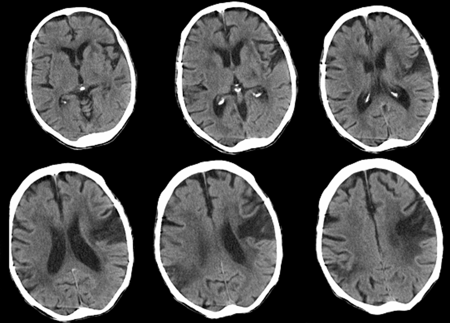
Changes in the rate of atrophy progression can be useful in diagnosing Alzheimer disease. Longitudinal changes in brain size are associated with longitudinal progression of cognitive loss, and enlargement of the third and lateral ventricles is greater in patients with Alzheimer disease than in control subjects.
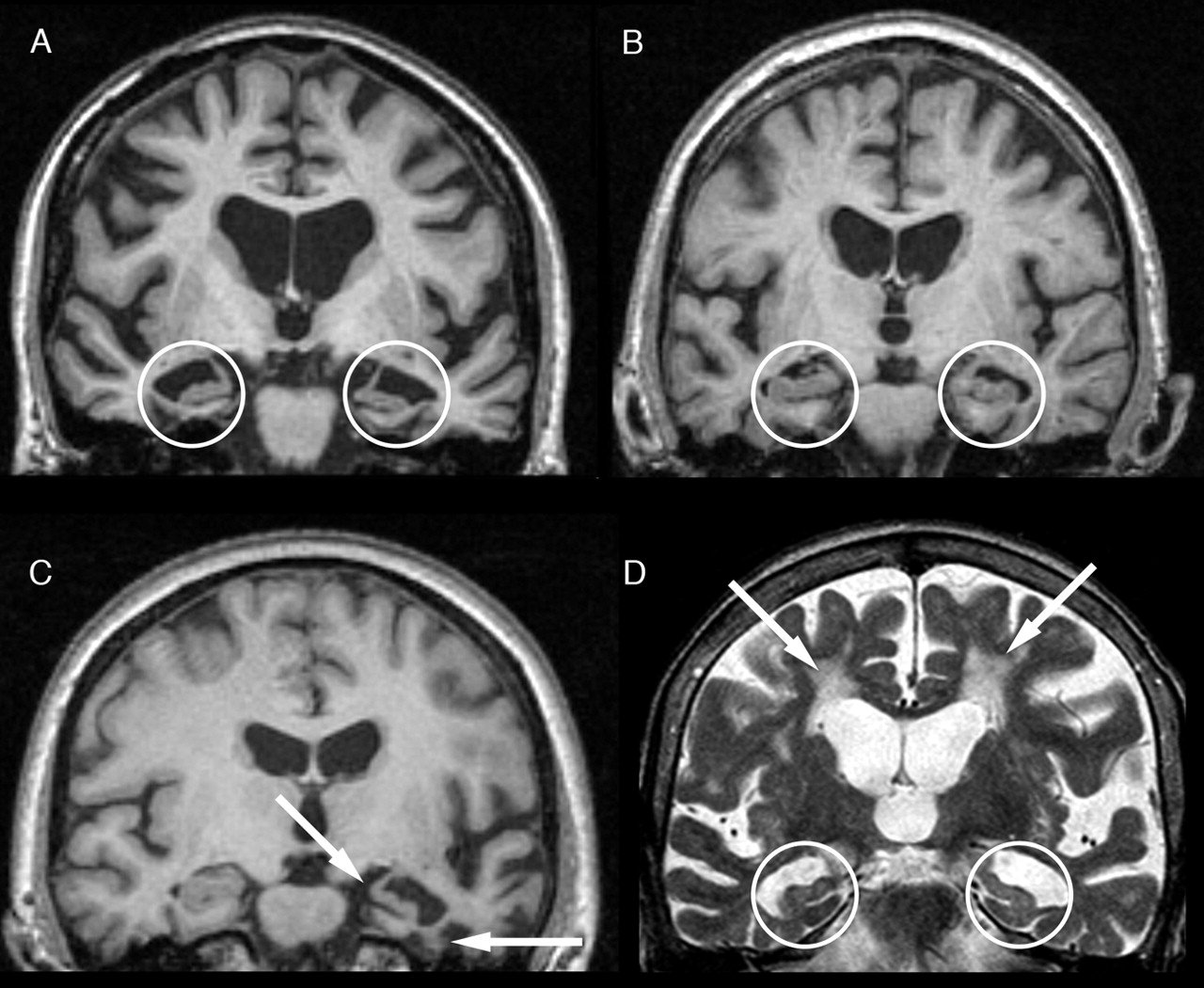
CT scan indices of hippocampal atrophy are highly associated with Alzheimer disease, but the specificity is not well established. Use of a nonquantitative rating scale showed a sensitivity of 81% and a specificity of 67% in differentiating 21 patients with Alzheimer disease with moderate dementia from 21 age-matched control subjects. Hippocampal volumes in a sample of similar size permitted correct classification of 85% of control subjects.
When Doctors Prescribe Brain Scans
Your physician might suggest that you get a brain scan to identify underlying problems causing mental conditions or affecting your general wellbeing.
Typically they are used to detect tumors, strokes, as well as other problems THAT CAN spark dementia that may appear on brain scans.
The cortex of the brain appears overly wrinkled and it has gyri which are separated by sulci .
Individuals with cortical atrophy experience the progressive loss of neurons which in turn causes the thinning of the ridges and the sulci to grow wider.
When brain cells continue dying, the brains fluid-filled cavities expand and occupy the available space.
In turn, they become LARGER than normal.
These structural changes within the brain are also aspects that BRAIN SCANS CAN IDENTIFY.
> > > Best Memory Loss Solution Available
If youre experiencing memory loss, you should go to a doctor. Your doctor will perform a physical exam and ask about your symptoms. He or she will also ask you about your medication and any stress youre experiencing. After the exam, he or she will likely ask you to make an appointment with a neuropsychologist. If youre unable to recall the details of your doctor, you may want to consult another healthcare provider.
Don’t Miss: What Are The Signs Of Frontotemporal Dementia
Understanding The Biology Of Ad
Importantly, imaging has a major role to play in improving our understanding of this disease . Uniquely, imaging is able to delineate in life the location within the brain of the effects of AD. Together with this topographical information imaging can quantify multiple different aspects of AD pathology and assess how they relate to each other and how they change over time. The clinical correlations of these changes and their relationships to other biomarkers and to prognosis can be studied. Ultimately the role of imaging in improving our understanding of the biology of AD underpins all its applications and is a theme that runs through the following sections of this article.
Initial Causes Ct Scan Of Normal Brain Vs Alzheimers

There are several different causes of memory loss. Some cause this condition in the young, while others may be more gradual. If you notice that your memory is weakening, its important to consult a medical professional. Whether the cause is mental illness, age, or a combination of factors, its important to seek treatment as soon as possible. People with extensive memory loss may have social difficulties and anxiety, which can lead to depression. They may be afraid they are letting their loved ones down, which can lead to anxiety and depression. Ct Scan of Normal Brain vs Alzheimers
Fortunately, there are many causes of memory loss, and many of them are treatable. However, if you are experiencing serious memory problems, you may need medical treatment. If you have been undergoing any type of medication, you should consult with your doctor. Some people have other underlying conditions that may be causing their loss of memory. Alcohol abuse, sleep deprivation, or other mental health conditions can cause memory problems. You should seek out a medical professional if you suspect youre suffering from any of these conditions.
Also Check: How Does A Person Die From Alzheimer’s Disease
Can A Ct Scan Show Dementia Conclusion
The bottom line is that CT scans and other brain imaging procedures CAN HELP diagnose dementia at any stage.
Combined with other assessments available, an individual battling the condition can get help early enough to manage it.
Families of people with dementia and caregivers can also access crucial information from these tests. They help in the process of caring for the individual.
Promoting Early Diagnosis Of Dementia
The early symptoms of dementia can include memory problems, difficulties in word finding and thinking processes, changes in personality or behaviour, a lack of initiative or changes in day to day function at home, at work or in taking care of oneself. This information does not include details about all of these warning signs, so it is recommended that you seek other sources of information. If you notice signs in yourself or in a family member or friend, it is important to seek medical help to determine the cause and significance of these symptoms.
Obtaining a diagnosis of dementia can be a difficult, lengthy and intensive process. While circumstances differ from person to person, Dementia Australia believes that everyone has the right to:
- A thorough and prompt assessment by medical professionals,
- Sensitive communication of a diagnosis with appropriate explanation of symptoms and prognosis,
- Sufficient information to make choices about the future,
- Maximal involvement in the decision making process,
- Ongoing maintenance and management, and
- Access to support and services.
You May Like: Why Do People Die From Alzheimer’s
Gray Matter Extraction From Brain Ct
We changed many default setting to the segmentation program in SPM8 taking the difference of CT and MR into account. Before using the segmentation function in SPM8, MRIcro and Image J were used to preprocess the CT images. The Brain Extraction Tool in MRIcro was used to remove the head holder segment. Image J was used to make the bounding box and voxel sizes equivalent to the tissue probability maps in SPM8. In SPM8, we set the segmentation parameters with extremely heavy regularization for unbiased CT images, a larger warp frequency cut-off of 35 Hz, a shorter sampling distance of 2, and a customized number of Gaussians per tissue class for each patients: 1 or 2 for gray and white matter and 68 for cerebrospinal fluid and other tissues. The number of Gaussians per tissue class was adjusted for each patient until successful segmentation was achieved. The CT images were then segmented to gray matter, white matter, cerebrospinal fluid, and other compartments using an unmodified version of the clustering algorithm .
Figure 1
Signs You May Need A Head Ct Scan
Some people with a family history of Alzheimers disease or dementia get a scan proactively for peace of mind. Others may have the scan after showing these symptoms of Alzheimers disease:
- Behavioral changes
- Difficulty understanding others when they speak
- Loss of appetite
- Mood changes, including irritability and depression
- Problems with depth perception
Memory loss is not inevitable with aging. If your loved one has memory loss and any of the above symptoms, it may be time to speak with your doctor about further testing.
Read Also: How Fast Does Alzheimer’s Progress
Why Have A Pet Scan
In the early 2000s, there was an exciting development it became possible to see amyloid in the brains of living people.
Scientists developed a special compound that could be injected into the bloodstream and attaches to clumps of amyloid protein in the brain. The compound emits a small radioactive signal that can be detected by a Positron Emission Tomography scanner.
Recognizing The Signs & Symptoms Of Alzheimer’s
Getting the correct scan to help detect Alzheimers is not the most challenging issue with this disease, the real challenge is recognizing the symptoms that indicate you may need to get a medical check-up and a diagnosis.
Its easy to ignore the early symptoms of the disease or, worse, not recognize the symptoms.
For example, after a certain age everyone starts to experience memory problems. We have all complained about going into a room and forgetting why we are there. However, when those incidents happen more often, or when you find yourself forgetting something that just happened a few minutes ago, that could be the sign of a deeper issue.
Friends and family members can also have problems recognizing that something is wrong.
For example, Alzheimers can cause personality changes. If the person goes from being sweet and easygoing to depressed and aggressive, you would probably notice. However, if the person goes from being depressed and aggressive to more depressed and aggressive, this is a more subtle change, and you might not realize that anything unusual is happening.
This is why it is important to know the common signs and symptoms of Alzheimer’s , so that you may be more likely to notice changes and discuss them with your doctor. The signs and symptoms of Alzheimer’s may include:
Having any or all of these symptoms could indicate Alzheimers or some other brain disorder, and should be discussed with your doctor.
Also Check: Can A Person Die From Dementia
What Can Brain Scans Tell Us About Dementia
The two characteristic changes in the brain of people who have Alzheimers disease are clumps of toxic proteins called amyloid and tau. Build-up of these proteins along with the death of brain cells defines the disease and leads to peoples symptoms.
The first account of these changes in the brain being related to the symptoms of Alzheimers disease is attributed to Alois Alzheimer in the early 1900s. The disease was later named after him. Seventy years later, the first computerised tomography brain scans were developed that revealed shrinkage of the brain due to loss of brain cells in people living with Alzheimers.
Since then, tools for measuring the size and shape of the brain have improved dramatically. Magnetic resonance imaging brain scans acquire beautifully detailed images of the structure of the brain, and can pinpoint more subtle areas of shrinkage as groups of brain cells die.
Until recently, spotting the toxic proteins was impossible except under the microscope, making it difficult to be certain whether the shrinkage was caused by Alzheimers disease, or one of the other less common causes of dementia.
Limitations Of Fmri In Ad

There are multiple challenges in performing longitudinal fMRI studies in patients with neurodegenerative dementias. It is likely that fMRI will remain quite problematic in examining patients with more severe cognitive impairment, as these techniques are very sensitive to head motion. If the patients are not able to adequately perform the cognitive task, one of the major advantages of task fMRI activation studies is lost. Resting state fMRI may be more feasible in more severely impaired patients.
It is critical to complete further validation experiments. BOLD fMRI response is known to be variable across subjects, and very few studies examining the reproducibility of fMRI activation in older and cognitively impaired subjects have been published to date . Longitudinal functional imaging studies are needed to track the evolution of alterations in the fMRI activation pattern over the course of the cognitive continuum from preclinical to prodromal to clinical AD. It is also important to evaluate the contribution of structural atrophy to changes observed with functional imaging techniques in neurodegenerative diseases. Finally, longitudinal multimodality studies, including structural MRI, fMRI, and FDG-PET and PET amyloid imaging techniques, are needed to understand the relationship between these markers, and the relative value of these techniques in tracking change along the clinical continuum of AD .
Read Also: What Happens If You Have Alzheimer’s Disease
Image Analysis Of 11c
Data analyses of 11C-PIB PET were performed using the PMOD software package . Distribution volume ratio images referenced to the cerebellum were generated using noninvasive Logan graphical analysis . Two experts in neuro-nuclear medicine, both with over 10 years of experience, interpreted the regional amyloid load, focusing on whether it was consistent with a diagnosis of AD.
What Are The Benefits Of Early Diagnosis
Early planning and assistanceEarly diagnosis enables a person with dementia and their family to receive help in understanding and adjusting to the diagnosis and to prepare for the future in an appropriate way. This might include making legal and financial arrangements, changes to living arrangements, and finding out about aids and services that will enhance quality of life for people with dementia and their family and friends. Early diagnosis can allow the individual to have an active role in decision making and planning for the future while families can educate themselves about the disease and learn effective ways of interacting with the person with dementia.
Checking concernsChanges in memory and thinking ability can be very worrying. Symptoms of dementia can be caused by several different diseases and conditions, some of which are treatable and reversible, including infections, depression, medication side-effects or nutritional deficiencies. The sooner the cause of dementia symptoms is identified, the sooner treatment can begin. Asking a doctor to check any symptoms and to identify the cause of symptoms can bring relief to people and their families.
Recommended Reading: Is There A Cure For Alzheimer’s 2017
Current Practice In Diagnosing Dementia
The remainder of this information will provide an overview of the diagnosis process and a guide to what happens after diagnosis.
It is important to remember that there is no definitive test for diagnosing Alzheimers disease or any of the other common causes of dementia. Findings from a variety of sources and tests must be pooled before a diagnosis can be made, and the process can be complex and time consuming. Even then, uncertainty may still remain, and the diagnosis is often conveyed as possible or probable. Despite this uncertainty, a diagnosis is accurate around 90% of the time.
People with significant memory loss without other symptoms of dementia, such as behaviour or personality changes, may be classified as having a Mild Cognitive Impairment . MCI is a relatively new concept and more research is needed to understand the relation between MCI and later development of dementia. However, MCI does not necessarily lead to dementia and regular monitoring of memory and thinking skills is recommended in individuals with this diagnosis.
> > > 1 Crazy Morning Recipe To Stop Brain Disease
Certain medications can also affect memory. A lack of sleep and an impaired thyroid function can negatively affect memory. Some of these conditions can also lead to a decreased ability to remember events. In addition to these, natural aging can affect brain function, and may lead to a slowdown in memory. Although this symptom does not necessarily mean that youre losing your memory, it could indicate a problem with your cognitive ability. If you are suffering from either, a medical evaluation is necessary to determine if youre suffering from memory loss. Ct Scan of Normal Brain vs Alzheimers
In addition to aging, medications can affect memory. Certain antidepressants, anxiety medications, and sleep disorders can all affect memory. A persons mental health can also contribute to memory problems. In some cases, a persons mental state may be affected by the medication they are taking. Some untreated medical conditions can lead to deterioration of the brain and affect the ability to learn and remember. It is also important to see a medical professional if your symptoms persist even after youve stopped taking certain medications.
Don’t Miss: What Stage Of Dementia Is Paranoia
Direct Comparison Between Spect And Pet In Alzheimer’s Disease
The technical characteristics of PET determines an overall higher performance over SPECT, in particular its greater sensitivity and spatial resolution. Even though significant, the differences in spatial resolution between both modalities are not of great magnitude, with values of 34 mm for PET and 58 mm for SPECT. The spatial resolution of PET is limited to 12 mm due to the positron emission range, while SPECT has no theoretical limitations in this regard, which has led to sub-millimeter resolutions in small animal equipment, exceeding the spatial resolution of their PET analogs. Dedicated brain SPECT cameras have the same spatial resolution as PET, although their availability is very limited and there has been no significant expansion of its use in clinical practice.
A critical question is to what extent the technical differences between both modalities influence the clinical diagnosis of neurodegenerative diseases, that is, whether or not they translate into considerable differences in diagnostic performance. The answer requires a critical review of the available literature. Considering systematic reviews or meta-analyses of the diagnostic value of both techniques in AD, Dougall et al. reported a sensitivity of 77% and specificity of 89% for SPECT while Patwardhan et al. including studies from the same time period reported a 86% sensitivity and specificity for PET . These results indicate a higher sensitivity and slightly lower specificity for PET.
Data Extraction And Quality Assessment
Data extraction and quality assessment were completed by one reviewer and checked by a second disagreements were resolved through discussion or referral to a third reviewer. We extracted data on: inclusion/exclusion criteria, included patients, CT and MRI technical and operator details, reference standard, imaging finding, definition of a positive imaging finding, numbers of patients in each patient group , and number of patients with positive imaging findings in each group. The patient groups were dichotomised as VaD or mixed dementia compared to AD or other diagnoses. This allowed construction of 2×2 tables of test performance, separately for each imaging finding assessed. Study quality was assessed using the Cochrane Collaborations adaption of the QUADAS tool .
Recommended Reading: How To Engage With Dementia Patients
Alternatives To A Head Ct Scan
Head CT scans may be the most effective way to diagnose Alzheimers disease. But if you prefer another method, magnetic resonance imaging of the head shows your doctor if you have mild cognitive impairment or brain shrinkage. Cognitive tests are available online to download, print and bring to a doctors office for a score. One option is the SAGE test from the Ohio State University Wexner Medical Center.
To get the most accurate diagnosis, your doctor may recommend combining tests and scans. For example, you can have a head MRI along with an assessment by your primary care doctor.
Facing the idea that you or a loved one might have Alzheimers disease is difficult. Its important though to not delay important imaging scans. Learn more about how CT scans help you and how to prepare for an upcoming appointment.