Basics Of Amyloid Pet As It Is Applied To Ad
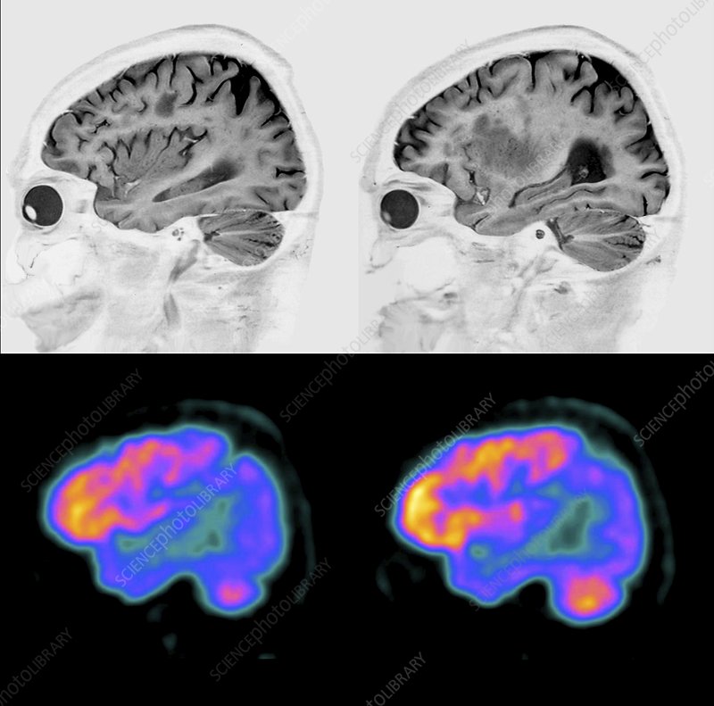
With regard to specific amyloid imaging agents, this review will discuss amyloid tracers in general, while acknowledging that most of the statements are derived from data on the most widely evaluated PET tracer, PiB . At the time of writing, there have been one or two, small published studies using each of the fluorine-18-labelled tracers, florbetaben , florbetapir and flutemetamol in AD patients. Although the PiB PET findings may ultimately be found to extend to these F-18-labeled tracers as well, this cannot be assumed until appropriate studies have been repeated with each individual tracer or until pharmacological equivalency to PiB has been established by direct comparison in the same subjects.
When You Need A Brain Scanand When You Dont
It is normal to forget things as you age. But many older people worry that they are getting Alzheimers disease when they cant remember things.
A new drug, used with a PET scan of the brain, can help diagnose Alzheimers. But before getting this scan you should have a complete medical exam. If your exam shows serious memory loss and your doctor cannot find a cause for it, then you should have the scan. Otherwise, the results can be misleading and you should not get the scan. Heres why:
The scan does not prove that you have Alzheimers.
Alzheimers can be found in the brain because it involves abnormal cell clumps. These clumps are called plaques. A PET scanwhich is an imaging testcan show these plaques, using a radioactive drug. During the test, the drug is injected into your body, where it attaches to the plaques. Then pictures are taken of your brain. The drug highlights the plaques so they can be seen on the scan.
If the scan does not show any plaques in your brain, then it is much less likely that you have Alzheimers. However, you can have plaques in your brain but not have Alzheimers. And having plaques does not mean that you will get Alzheimers in the future.
Alzheimers is not the only cause of forgetting things.
Medicines can also cause memory loss and thinking problems. So if you have symptoms, it is important to find out what the cause is.
Finding the cause starts with a medical evaluation.
The new scan can pose risks.
It can be expensive.
02/2013
Brain Scans Show Risk Factor For Alzheimer’s
Changes in Brain Chemistry Linked With Lower Scores on Memory, Language Tests
In the study, researchers used a special MRI scan and a special PET scan in people in their 70s and 80s who were aging normally to help identify those who had brain changes thought to be linked with Alzheimer’s disease.
The scans looked for amyloid-beta plaques, one of the early changes linked with the disease, and for biochemical changes, says researcher Kejal Kantarci, MD, associate professor of radiology at the Mayo Clinic in Rochester, Minn.
About a third had high levels of plaques, she found. Those who had high levels of plaques on the PET scan also tended to have the biochemical changes found on the MRI.
“We found that these biochemical changes in the brain of normally aging people were associated with worse performance on tests of mental abilities, including memory, language, and attention,” she tells WebMD.
The approach is one of several under study to identify those most at risk for developing Alzheimer’s disease. Others are working on blood tests, for example.
The most common form of dementia, Alzheimer’s affects about 5.4 million Americans, according to the Alzheimer’s Association. While doctors often use brain imaging, evaluation of behavior, psychiatric tests, and other means to diagnose the disease, none is highly accurate.
The new study was funded by the National Institutes of Health. It is published in Neurology.
Read Also: Does Diet Coke Cause Memory Loss
Ct Scans: How They Help Detect Alzheimers Disease
Early diagnosis of Alzheimers disease allows you to treat it sooner and stay healthy and independent for longer. Head CT scans are one the most effective tools to detect the condition. Learn how a head CT scan can help you or your loved one with memory loss determine if an advanced condition like Alzheimers disease is the root of the symptoms.
The Mri Scan: How Can It Be Used In The Diagnosis Of Alzheimer’s

As medicine has improved in the last few decades, the role of these brain imaging studies has become more and more important in the diagnosis of the Alzheimer’s Disease and other neurological conditions. One of the key diagnostic tests is known as Magnetic resonance imaging the MRI.
In laymen’s terms, an MRI scan is an imaging study that relies on a magnetic field in order to get a good view of your internal organs. The magnetic field can actually influence the water molecules that are naturally found in your body in order to give a very refined picture of what is going on in your body. It is best to use this type of imaging for soft tissue the exact type of tissue that you will be looking at when you are investigating the brain.
An MRI scan can be a rather long examination for patients to endure and patients will need to lie as still as possible in order for doctors to get an accurate picture of the structures being investigated. During just a simple head investigation, patients may be asked to lie still for at least 30 minutes. This may sound like a simple task but the tubing in the room is rather small so many patients may not enjoy the slight claustrophobia that can easily occur. It is one of the best studies to do, though, so the accurate results are worth the slight discomfort for the patient.
You May Like: Smelling Farts Can Prevent Cancer
Rate Of Change Of Brain Atrophy
Changes in the rate of atrophy progression can be useful in diagnosing Alzheimer disease. Longitudinal changes in brain size are associated with longitudinal progression of cognitive loss, and enlargement of the third and lateral ventricles is greater in patients with Alzheimer disease than in control subjects.
CT scan indices of hippocampal atrophy are highly associated with Alzheimer disease, but the specificity is not well established. Use of a nonquantitative rating scale showed a sensitivity of 81% and a specificity of 67% in differentiating 21 patients with Alzheimer disease with moderate dementia from 21 age-matched control subjects. Hippocampal volumes in a sample of similar size permitted correct classification of 85% of control subjects.
Basics Of Functional Mri As Applied To Ad
Functional MRI is being increasingly used to probe the functional integrity of brain networks supporting memory and other cognitive domains in aging and early AD. fMRI is a noninvasive imaging technique which provides an indirect measure of neuronal activity, inferred from measuring changes in blood oxygen leveldependent MR signal . Whereas fluoro-deoxy-d-glucose -PET is thought to be primarily a measure of synaptic activity, BOLD fMRI is considered to reflect the integrated synaptic activity of neurons via MRI signal changes because of changes in blood flow, blood volume, and the blood oxyhemoglobin/deoxyhemoglobin ratio . fMRI can be acquired during cognitive tasks, typically comparing one condition to a control condition , or during the resting state to investigate the functional connectivity within specific brain networks. Fc-MRI techniques examine the correlation between the intrinsic oscillations or time course of BOLD signal between brain regions , and have clearly documented the organization of the brain into multiple large-scale brain networks . Both task-related and resting fMRI techniques have the potential to detect early brain dysfunction related to AD, and to monitor therapeutic response over relatively short time periods however, the use of fMRI in aging, MCI, and AD populations thus far has been limited to a relatively small number of research groups.
Read Also: What Color Represents Alzheimer’s
Utility Of Fdg Pet In The Study Of Ad
The Pattern of FDG Hypometabolism Is an Endophenotype of AD
A substantial body of work over many years has identified a FDG-PET endophenotype of AD that is, a characteristic or signature ensemble of limbic and association regions that are typically hypometabolic in clinically established AD patients . The anatomy of the AD signature includes posterior midline cortices of the parietal and posterior cingulate gyri, the inferior parietal lobule, posterolateral portions of the temporal lobe, as well as the hippocampus and medial temporal cortices. Metabolic deficits in AD gradually worsen throughout the course of the disease. Bilateral asymmetry is common at early stages, more advanced disease usually involves prefrontal association areas, and in due course even primary cortices may be affected. Interestingly, the regions initially hypometabolic in AD are anatomically and functionally interconnected and form part of the large-scale distributed brain network known as the default mode network . We now know in addition that these regions are highly vulnerable to amyloid- deposition .
FDG Hypometabolism Is Related to Other AD Biomarkers and to Genes
FDG PET Is a Valid AD Biomarker
Brain Scans Spot Track Alzheimer’s
HealthDay Reporter
TUESDAY, April 2, 2019 — Brain scans can improve diagnosis and management of Alzheimer’s disease, a new study claims.
Researchers assessed the use of PET scans to identify Alzheimer’s-related amyloid plaques in the brain. The study included more than 11,000 Medicare beneficiaries with mild thinking impairment or dementia of uncertain cause.
This scanning technique changed the diagnosis of the cause of mental impairment in more than one-third of the participants in the study.
The brain scan results also changed management — including the use of medications and counseling — in nearly two-thirds of cases, according to the study published April 2 in the Journal of the American Medical Association.
“These results present highly credible, large-scale evidence that amyloid PET imaging can be a powerful tool to improve the accuracy of Alzheimer’s diagnosis and lead to better medical management, especially in difficult-to-diagnose cases,” said study co-author Maria Carrillo, chief science officer of the Alzheimer’s Association.
“It is important that amyloid PET imaging be more broadly accessible to those who need it,” she added in an association news release.
Funding for the study came from Avid Radiopharmaceuticals Inc., General Electric Healthcare, and Life Molecular Imaging.
There is no cure for Alzheimer’s disease, but early diagnosis means that patients can receive treatment to manage symptoms and be directed to clinical trials for new drugs.
Continued
Also Check: How Fast Does Ftd Progress
Current Practice In Diagnosing Dementia
The remainder of this information will provide an overview of the diagnosis process and a guide to what happens after diagnosis.
It is important to remember that there is no definitive test for diagnosing Alzheimers disease or any of the other common causes of dementia. Findings from a variety of sources and tests must be pooled before a diagnosis can be made, and the process can be complex and time consuming. Even then, uncertainty may still remain, and the diagnosis is often conveyed as possible or probable. Despite this uncertainty, a diagnosis is accurate around 90% of the time.
People with significant memory loss without other symptoms of dementia, such as behaviour or personality changes, may be classified as having a Mild Cognitive Impairment . MCI is a relatively new concept and more research is needed to understand the relation between MCI and later development of dementia. However, MCI does not necessarily lead to dementia and regular monitoring of memory and thinking skills is recommended in individuals with this diagnosis.
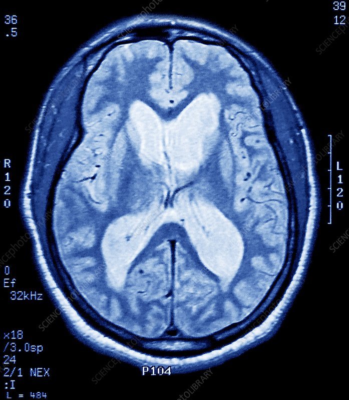
Magnetic Resonance Imaging Brain Scan
MRIs are often located in radiology centers or hospitals. MRIs work by aligning the hydrogen molecules in your cells using powerful magnets. As the molecules absorb the energy from the radiofrequency waves, they emit a signal in response. With software help, the data becomes a detailed image. Bodily structures with high water content, like the soft tissues and organs such as the brain and kidneys, create the most contrast and therefore produce better MRI images.
Because MRI machines use strong magnetic fields, this type of medical imaging isnt right for everyone. Let your provider know if you have any implanted medical devices and surgical hardware , dental implants, metal foreign bodies, tattoos, and permanent make-up.
If you are claustrophobic, let your health provider know. You may also inquire about using an open-MRI which, as its name suggests, is open on at least three sides. With no tunnel to fit into, open MRI scanners can accommodate more body types.
A major drawback of open MRIs is poor image quality. Because of its open-air design, the scanner loses some of its magnet strength. Instead of an open MRI, Ezra uses 3T MRI scanners at all of their partner facilities. These scanners feature a larger bore with a shorter tunnel, a stronger magnet, and faster scan times.
Don’t Miss: Alzheimer’s Disease Color Ribbon
Mri Scans Can Show Dementia
According to researchers from Perelman School of Medicine at the University of Pennsylvania, the answer to can an MRI detect dementia is to some extent yes.
The scientists explained that doctors have an easier time telling whether a person has dementia through MRI scans.
This gets rid of the need to carry out invasive tests that people find unfriendly like the lumbar puncture where a doctor must stick a needle in the spine.
Additionally, it also helps to speed up the diagnosis process which is important seeing that dementia diagnosis for the longest time has been a struggle for medics often leading to delayed treatment.
In addition to telling whether a person has dementia, MRI scans may in the future help doctors determine whether an individual is at risk of dementia according to new research.
Research from the University of California San Francisco and the Washington University School of Medicine in St. Louis conducted a small study where MRI brain scans were able to predict with 89% accuracy the people who were going to develop dementia in three years.
The researchers presented their findings in Chicago during a Radiological Society of North America meeting.
It suggested that in a few years, physicians will be able to tell people their risk of developing dementia before they start to showcase any symptoms of the neurodegenerative illness.
How Accurate Is Magnetic Resonance Imaging For The Early Diagnosis Of Dementia Due To Alzheimers Disease In People With Mild Cognitive Impairment

Why is improving Alzheimers disease diagnosis important?
Cognitive impairment is when people have problems remembering, learning, concentrating and making decisions. People with mild cognitive impairment generally have more memory problems than other people of their age, but these problems are not severe enough to be classified as dementia. Studies have shown that people with MCI and loss of memory are more likely to develop Alzheimers disease dementia than people without MCI . Currently, the only reliable way of diagnosing Alzheimers disease dementia is to follow people with MCI and assess cognitive changes over the years. Magnetic resonance imaging may detect changes in the brain structures that indicate the beginning of Alzheimers disease. Early diagnosis of MCI due to Alzheimers disease is important because people with MCI could benefit from early treatment to prevent or delay cognitive decline.
What was the aim of this review?
To assess the diagnostic accuracy of MRI for the early diagnosis of dementia due to Alzheimers disease in people with MCI.
What was studied in the review?
The volume of several brain regions was measured with MRI. Most studies measured the volume of the hippocampus, a region of the brain that is associated primarily with memory.
What are the main results in this review?
How reliable are the results of the studies?
Who do the results of this review apply to?
What are the implications of this review?
How up to date is this review?
Recommended Reading: How Fast Does Alzheimer’s Progress
What Is Alzheimers Disease
Alzheimers is thought to be the result of beta-amyloid plaques, which are thick protein deposits present in the brain, and neurofibrillary tangles abnormal structures, which form in neurons building up in the brain. This buildup causes neurons in the brain to cease working, losing connection with other neurons before dying. However, the exact cause of Alzheimers is still unknown. The condition is the most common form of dementia and can quickly progress from being mild to severe.
There are no known causes for Alzheimers since the disease is still being studied. A persons chances of developing Alzheimers tend to increase with age, but people in their 40s and 50s can begin showing symptoms of early-onset Alzheimers. People who have a relative with the condition may also be at higher risk of developing it themselves since the disease has hereditary factors.
A persons overall physical health may also be a factor in whether they develop Alzheimers disease. One study showed that people who have heart disease or other cardiovascular issues could be at a higher risk of developing Alzheimers than those with good heart health. This is because cardiovascular issues often reduce the amount of blood the brain receives, which may increase the cognitive issues associated with Alzheimers.
Read Also: Ribbon Color For Dementia
At Risk Of Dementia Brain Scan Shows When You Might Develop Symptoms Study Says
With just one brain scan and a persons age, an algorithm can reveal how much time is left plus or minus several years before someone at risk of developing dementia will begin to experience symptoms of the condition.
The Washington University School of Medicine study found a correlation of 0.9 between the age someone is expected to first show symptoms and the true age of diagnosis . The study was .
The technique uses data from amyloid positron emission tomography , a widely used brain scan in Alzheimers research, to measure the levels of the beta-amyloid protein in the brain. The algorithm offers a new way to analyze this data that helps researchers determine an estimated timeline of symptom onset.
Dementia is an umbrella term for a group of conditions that affect a persons ability to remember things or make decisions on a daily basis, with Alzheimers being the most common type. In 2014, there were an estimated 5 million adults at least 65 years old with dementia in the U.S., according to the Centers for Disease Control and Prevention. By 2060, that number is predicted to rise to nearly 14 million.
While some people may not want to know when theyll start to forget friends names or have difficulty calculating change at the grocery store, others, particularly those with genetic predispositions for dementia, could benefit from having time to prepare for the inevitable changes.
Recommended Reading: What Color Is Alzheimer’s Awareness Ribbon