Hyperexcitability And Seizures At Early Ad Stages
It has been reported that during memory tasks individuals with mild AD show reduced hippocampal activity, whereas aMCI and MCI patients exhibit hyperactivity in the hippocampus/parahippocampal region . Task-related hyperactivity has been described in asymptomatic carriers of AD pathological mutations during associative encoding task ; in asymptomatic offspring of autopsy-confirmed AD patients ; in cognitively intact young and old carriers of APOE4 and in low-performing clinically healthy aged individuals . Conversely, individuals at late aMCI stage and early AD already express the hippocampal hypoactivity pattern . During an associative memory task hyperactivation of the anterior hippocampus and EC in MCI patients compared to the controls was observed . Recently, the characteristic pattern of hyperactivity as well as shape and volume changes were detected in the CA3/DG of aMCI patients . CA3/DG network is essential for a key process for episodic memory, pattern separation .
The occurrence of seizures in AD might have serious implications for neurogenesis. Seizures have been shown to increase neurogenesis, however they eventually decrease the survival of new-born neurons which also show aberrant migration and form aberrant circuits . Recently, Hester and Danzer demonstrated that status epilepticus similarly affects both dorsal and ventral hippocampus integration of new-born granule neurons and mossy fiber sprouting .
Nlgn1 Deletion Reveals Neuronal Loss After Treatment With Ao1
Lastly, we asked whether the absence of NLGN1 impacts neuronal loss in the DG caused by chronic hippocampal Ao1-42 injections. This was assessed using the same mice as above tested for spatial and working memory. Neuronal loss was measured in the DG in an area located under the injection site . The size of the counting area did not differ between groups . However, DG neuronal count was significantly decreased in Nlgn1 KO mice submitted to Ao1-42 injections in comparison to all other groups . Of note is that WT mice injected with Ao1-42 did not significantly differ from WT mice with control injections, thus confirming an intermediate exposure to Ao1-42. Neuronal count in a region distant to the injection site revealed no significant difference between the four groups . These results suggest that the absence of NLGN1 potentiates the toxicity of Ao1-42, leading to a noticeable neuronal loss in the DG.
Figure 6
What Happens If Hippocampus Get Damaged
The Hippocampus is especially sensitive to the global reductions in the level of oxygen in the body. So, periods of oxygen deprivation or Hypoxia, which are not fatal, may nonetheless, cause particular damage to the hippocampus. This could occur, usually during a heart attack, respiratory failure, carbon monoxide poisoning, and sleep apnea.
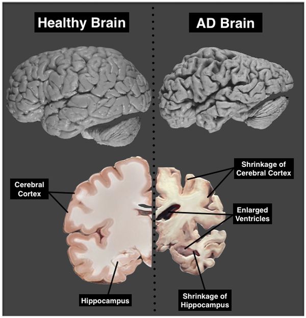
Damaged Hippocampus can cause loss of memory and difficulty in establishing new memories. In Alzheimers disease, the Hippocampus is one of the first regions of the brain that is affected, leading to the confusion and loss of memory, that is so commonly seen in the early stages of the disease. This small brain structure may also be damaged through chronic seizures in epilepsy. Even, Encephalitis can cause a damage to the Hippocampus. Schizophrenia, is one more condition that may be associated with a damage to the Hippocampus.
One symptom of damage to the Hippocampus is Amnesia, or the loss of some portion of the memory. Apart from this, a damage to the Hippocampus can also cause poor impulse control, hyperactivity, and difficulty with spatial navigation or memory.
Real-life amnesia, generally does not mean that you lose your sense of identity. In fact, the individuals know who they are, however, may have trouble learning new facts and forming new memories. Though there are no specific treatments for Amnesia, there are techniques for enhancing memory and even psychological support can help the patients
Also Check: Which Of The Following Symptoms Is Suggestive Of Alzheimer Disease
Impact Of Hippocampus Damage
If the hippocampus is damaged by disease or injury, it can influence a person’s memories as well as their ability to form new memories. Hippocampus damage can particularly affect spatial memory, or the ability to remember directions, locations, and orientations.
Because the hippocampus plays such an important role in the formation of new memories, damage to this part of the brain can have a serious long-term impact on certain types of memory. Damage to the hippocampus has been observed upon post-mortem analysis of the brains of individuals with amnesia. Such damage is linked to problems with forming explicit memories such as names, dates, and events.
The exact impact of damage can vary depending on which hippocampus has been affected. Research on mice suggests that damage to the left hippocampus has an effect on the recall of verbal information while damage to the right hippocampus results in problems with visual information.
If Your Hippocampus Is Bigger Than Normal Don’t Worry About Getting Alzheimer’s

More volume, more memory may sound like the tagline for a new hair commercial but, in fact, its the nutshell results of a preliminary study which suggests;the bigger the size of your hippocampus, the lower your risk of memory decline and dementia.
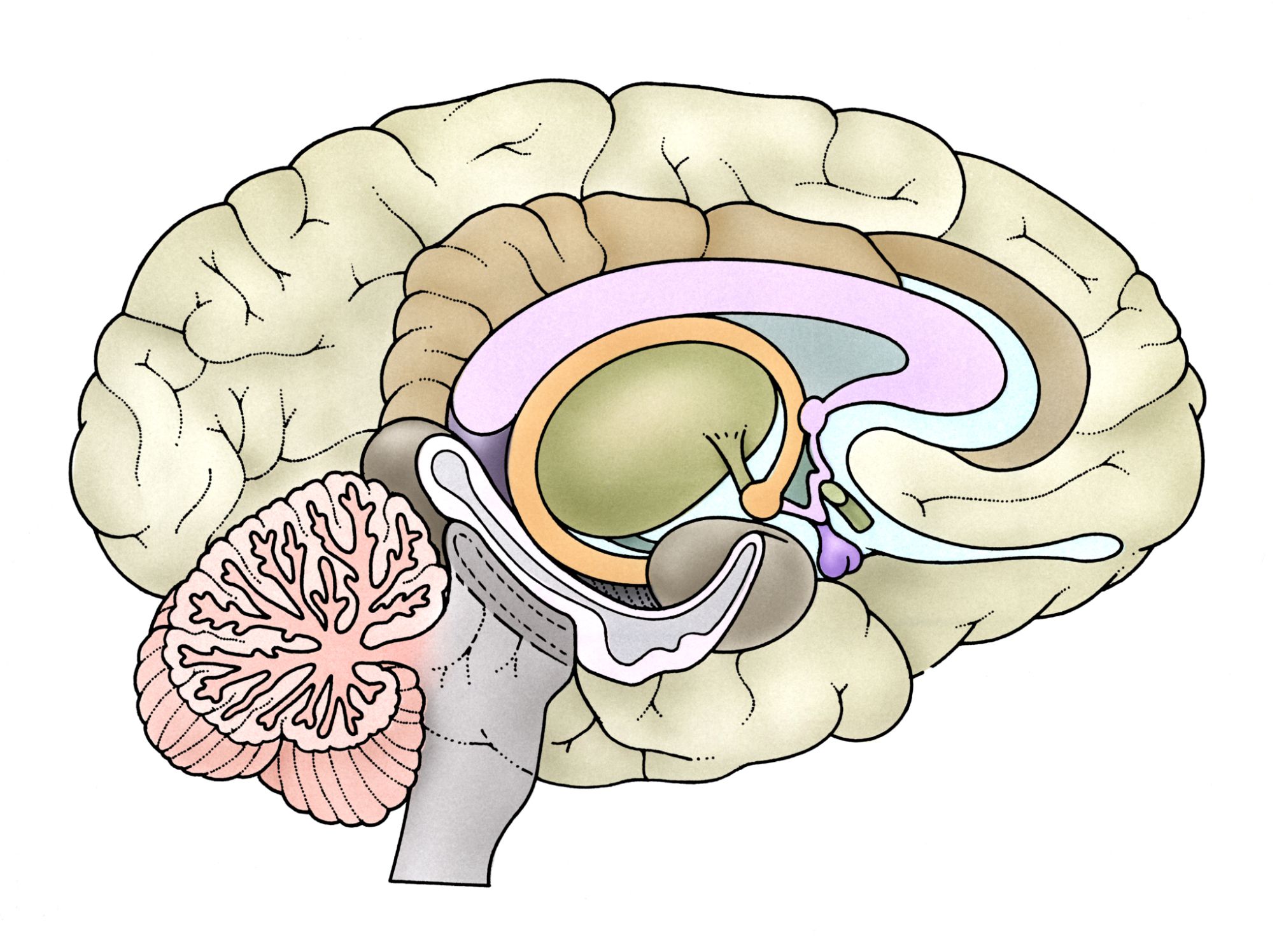
The two seahorse-shaped structures tucked deep inside the brain on both sides, left and right, are referred to as the hippocampus. As part of the limbic system, the hippocampus plays a role in both emotion and the formation of new memories. Specifically, the hippocampus is where information usually emotionally-charged information is transferred, like some hasty transaction performed at the ATM, into the long-term memory banks. While the left hippocampus appears to help us retain words and language, the right hippocampus is linked to spatial memory, such as the layout of streets in your hometown.
Unsurprisingly, the hippocampus is one of the first structures to erode in the brains of patients with Alzheimer’s disease, a type of dementia that causes problems with memory, thinking, and behavior. Many previous studies of dementia have focused on the hippocampus and for the current study, the research team investigated how the size of this brain structure relates to risk of Alzheimer’s disease.
Led by Aaron Bonner-Jackson, the team examined 226 patients at the Center for Brain Health, Cleveland Clinic, to search for indications of dementia.
Recommended Reading: How To Differentiate Delirium From Dementia
Does The Hippocampus Play A Role In Determining Ptsd Risk
Not everyone who experiences a traumatic event develops PTSD. Therefore, researchers have also proposed that the hippocampus may play a role in determining who is at risk for developing PTSD.
Specifically, it is possible that having a smaller hippocampus may be a sign that a person is vulnerable to developing a severe case of PTSD following;a traumatic event. Some people may be born with a smaller hippocampus, which could interfere with their ability to recover from a traumatic experience, putting them at risk for developing PTSD.
In twin studies that focused on;identical twins, with one twin exposed to a traumatic event and the other unexposed, researchers are able to look at pre-existing vulnerabilities that may be present in both twins, as well as differences that may be due to trauma. Since twin participants share the same genes, studying identical twins can provide insight into the influence of genetics on developing certain conditions.
For example, in this case, if the person who developed PTSD has a smaller hippocampus and has a non-trauma exposed twin who has a smaller hippocampus, it would suggest that a smaller hippocampus may be a sign of genetic vulnerability for developing PTSD following a traumatic experience.
What Happens When The Hippocampus Is Damaged
In patients with Alzheimer’s disease,;one of the first things to falter is the ability to make new memories because of the gradual decrease in size of the hippocampus, according to a 2012 review published in the journal Annals of Indian Academy of Neurology. The gradual decline in size and function of this part of the brain is also associated with a string of other severe mental illnesses, such as depression, schizophrenia and epilepsy.;
According to Epilepsy Research UK, hippocampus damage has been observed in 50-75% of patients with epilepsy who had autopsies,;but it is not yet clear that the damage is a cause or consequence of recurrent seizures.;
In general, the hippocampus is a particularly vulnerable part of the brain and can be adversely affected by many different conditions, including long-term exposure to high levels of stress, or head injury, the 2012 review concluded.;
Recommended Reading: How Early Can Dementia Start
Morris Water Maze Test
The MWM task was conducted 48hours after the Y-maze test as previously described. Mice were trained in a 120-cm diameter pool filled with water rendered opaque by adding non-toxic white paint. The temperature of the water was kept at 24±1°C during testing. During the first 4 days, mice had to learn to find a submerged platform in the pool using visuo-spatial cues installed around the pool. Mice had 3 trials per day with 30min between each trial. If the mouse failed to reach the platform within 60sec, it was guided to the platform and had to remain on the platform for 10sec before being removed from the pool. The time and distance to reach the platform were extracted by the Smart software . Starting location of the mice was different for each trial. During the probe trial on the fifth day, the platform was removed and mice had 60sec to explore the pool. The time spent in the target quadrant that previously contained the platform and in the other quadrants, as well as the number of times the mouse crossed the area around the platform were calculated with the Smart software. A final cued task of 3 trials was also performed to verify visual acuity and motivation to escape from water. During this test, mice had a maximum of 30sec to reach a visible platform that was moved in a different quadrant between each trial. Data were acquired in real time with the Smart software.
What Happens To The Brain In Alzheimer’s Disease
The healthy human brain contains tens of billions of neuronsspecialized cells that process and transmit information via electrical and chemical signals. They send messages between different parts of the brain, and from the brain to the muscles and organs of the body. Alzheimers disease disrupts this communication among neurons, resulting in loss of function and cell death.
Also Check: Why Do Alzheimer’s Patients Play With Their Feces
How Alzheimer’s Disease Affects The Hippocampus
Research has found that one of the first areas in the brain affected by Alzheimer’s disease is the hippocampus. Scientists have correlated atrophy of the hippocampal areas with the presence of Alzheimer’s disease. Atrophy in this area of the brain helps explain why one of the early symptoms of Alzheimer’s disease is often impairment of memory, especially the formation of new memories.
Hippocampus atrophy has also been correlated with the presence of tau protein that builds up as Alzheimer’s disease progresses.
Spatial Object Recognition Test
The SOR task was conducted 24hours after the last hippocampal injection in Nlgn1 KO mice and wild-type littermates similar to previously described. It was done in an opaque plastic arena with the floor covered with litter that was mixed after each trial to eliminate olfactory cues. The four walls contained different visual cues and two different objects were placed in adjacent corners of the arena. The walls and objects were cleaned with 70% ethanol between each mouse. During the first day, mice explored the arena and objects for 5min. Twenty-four hours later, one of the two objects was moved to the opposite corner of the arena and mice were reintroduced in the arena for 5min. The number of interactions with each object, defined as a touch of the snout or sniffing of the object by the mouse, was counted by an experimenter blind to the genotype and treatment. The discrimination index was calculated for the number of interactions according to the following formula: /.
Read Also: What Is The Average Lifespan Of A Person With Dementia
Relationship Between Ad Pathology In The Brainstem And Osa Severity
The frequency of no pathology, tau-only pathology, A-only pathology, and tau + A pathology can be seen in , separated into those with lower and higher ODI scores. The lower and higher ODI groups had similar numbers of people with each type of pathology. The percentage of brains containing any tau is similar for both lower and higher ODI . The percentage of brains containing any A is higher for those with higher ODI; however, only five brains contained A. A Fishers Exact Test for independence found that there was no significant difference between the distribution of pathology depending on OSA severity . Further, no significant correlations were found between log ODI and the number of NFTs in the brainstem overall, r2 = 0.053, p = 0.289, or the number of NFTs in the LC, r2 = 0.003, p = 0.789. However, the small number of brainstems with NFTs prevented us from performing regression analyses to control for age, sex, CPAP use, and BMI, so we cannot rule out the possibility that these factors are obscuring a relationship between ODI and NFT.
Percentage of subjects in the lower and higher ODI groups with tau and/or A pathology in the pons at the level of the LC
| .; |
|---|
Hippocampus Brain Injury: Key Points

The hippocampus is one of the most crucial structures of thebrain. Not only does it play a pivotal role in the formation of new memories,but it also helps the brain produce new nerve cells.
An injury to the hippocampus can cause serious memory problems. But fortunately, physical and cognitive exercises can help reverse some of the worst effects of hippocampal damage and improve your memory skills. ;
Also Check: Why Does Uti Cause Dementia In Elderly
Treatments For The Hippocampus
While many of the problems associated with hippocampus deterioration have no absolute cure, there are things you can do to keep your hippocampus healthy.
Maintain a healthy diet
Limit your intake of saturated fat and stick to healthy fats, whole grains, fruits, and vegetables. Look for foods that are rich with antioxidants. You should also consume foods with omega-3 fatty acids such as fish or olive oil. Avoid alcohol, which has been associated with hippocampus atrophy.
Stay mentally and socially active
Both mental and social stimulation are important to hippocampus size. Keep your brain healthy with puzzles, games, and new challenges. Social relationships and interaction also play a role. They help ward off depression, which is associated with hippocampal atrophy. Studies of older people routinely demonstrate the importance of social activity to hippocampus function.
Exercise
Exercise may help you keep the hippocampus healthy through both direct and indirect means. Its indirect benefits come from exercises ability to lower blood pressure and prevent metabolic issues, both of which are linked to brain decay.
Aerobic exercise is particularly important and directly benefits the hippocampus. One study found that regular aerobic exercise boosts the size of the hippocampus.
Stay current with medical developments
Talk to your doctor about recent developments in the field and get updates as necessary.
Hippocampal Asymmetry In Ad
Although hippocampus is structurally and functionally asymmetric, right vs. left hippocampal volume differences have received less research attention. In healthy adults there is hemispheric asymmetry of the whole hippocampus, with larger volume of the right one . There are also right vs. left differences in the layers thickness and volumes of different hippocampal subfields. For instance, Lister et al. identified asymmetries in neuronal numbers in rat CA1 and CA3/CA2 subfields, with the right hemisphere containing 21 and 6% fewer neurons, respectively .
Hippocampal volume asymmetry has been connected with cognitive functions and it has been suggested that hippocampal subfields analysis should be included in these correlation studies. For instance, Woolard and Heckers in a study of 110 healthy individuals of 32.3 ± 10.7 years of age demonstrated that the R > L asymmetry is limited to the anterior hippocampus and it is correlated with a measure of general cognitive functions . Moreover, they showed that the volume of anterior hippocampus correlates with the volumes of all four cortical lobes, whereas the posterior hippocampus volume was found strongly correlated with the volume of occipital cortex .
Read Also: How Do You Spell Dementia
Nlgn1 Level Is Decreased By Ao1
Given our observation that NLGN1 level significantly correlates with soluble A in the human hippocampus , we next verified whether Ao1-42 could directly impact NLGN1 level in vitro, and whether this is linked to neuronal survival. NLGN1 protein level and cell viability were measured in primary culture of hippocampal neurons treated with 2 M of Ao1-42 for 48 or 72hours. As expected from our previous study, Ao1-42 significantly decreased neuronal viability by about 40% after 72hours of treatment . No change in viability was observed after 48hours of treatment. NLGN1 protein level was also decreased after 72hours of Ao1-42 exposure and unchanged after 48hours . These data indicate that Ao1-42 exposure is detrimental to NLGN1 protein level in hippocampal neurons, which follows a time course similar to neuronal viability.
Figure 3
Relationship Between Nft Burden In The Hippocampus And Osa Severity
In the hippocampus sections, NFTs were observed in 24 of the 34 brains. Of these 24, 7 were at Stage 1, 12 were at Stage 2, 3 were at Stage 3, and 2 were at Stage 4 . Ordinal regression analysis was performed with NFT stage as the dependent variable . Log ODI was a significant predictor of NFT stage when it was the only predictor in the model . However, when age is included as a predictor in the model, NFT stage is no longer significant and age is a significant predictor . Age remains the only significant predictor when CPAP use, sex, and BMI are also added to the model .
You May Like: How Fast Can Vascular Dementia Progress