Possible Limitations And Future Directions
The four population-based studies reviewed here employed relatively crude measures of CAA. There were varying attempts to take into account the interactions with a wide range of other dementia-related pathologies and the severity of CAA. No study took into account the distribution of CAA, the presence of inflammation and the type of vessel affected. All these factors have been reported to influence the association with cognition ].
Population-based studies, as with studies on selected samples, face some important stumbling blocks in the investigation of CAA. The distribution of CAA is variable and its detection critically dependent of the extent and distribution of histological sampling. There is likely to be an under-diagnosis of CAA even in severe cases . There is currently no consensus as to how to sample for or detect CAA or grade its severity. Full evaluation of CAA is most satisfactorily assessed by isolating cerebral and leptomeningeal vessels from the brain and staining for amyloid with Thioflavin . A standard consensus method for detecting and classifying CAA would greatly facilitate future population-based multi-centre studies of CAA and such plans are underway.
Amyloid Protein And Hemorrhage
Amyloid damages the media and adventitia of cortical and leptomeningeal vessels, leading to thickening of the basal membrane, stenosis of the vessel lumen, and fragmentation of the internal elastic lamina. These processes result in fibrinoid necrosis and microaneurysm formation, predisposing to hemorrhage.
CAA-related brain changes include lobar cerebral and cerebellar hemorrhage, leukoencephalopathy, small cortical ischemic infarcts, and plaque deposition. Leukoencephalopathy may be related to chronic hypoperfusion of deep WM .
Neuropathologically, mild CAA primarily affects a relatively smaller proportion of the leptomeningeal and superficial cortical vessels, in contrast to the diffuse, significant deposition of amyloid in small arteries and arterioles seen in severe CAA. Medium-sized leptomeningeal arteries are affected with amyloid deposition in the outer portion of tunica media to tunica adventitia.
Frequently, complete erosion occurs, with only endothelium surrounding the deposit, predisposing to hemorrhage. Electron microscopy demonstrates fibrils of amyloid in the outer basement membrane in the initial stage of CAA. As the disease progresses, significant amyloid accumulation leads to tunica media degeneration, capillary and arteriolar infiltration, and formation of dystrophic neuritic plaques.
Convincing Effects Of Phosphodiesterase Inhibitor
Among varieties of vasoactive drugs, cilostazol, a selective inhibitor of type 3 phosphodiesterase , is likely to be a promising agent for AD and CAA . PDE3 can hydrolyze both cAMP and cGMP, while increasing cAMP level is a major pharmacological effect of cilostazol . PDE3 is widely expressed in central nervous system and up-regulated in A-positive vessels, especially in vascular smooth muscle cells , suggesting the possibility that PDE3 inhibition could be therapeutic for CAA. Cilostazol possesses multiple effects, such as increasing pulse rate and arterial elasticity , prolonging pulse duration time , and dilating cerebral vessels such vasoactive actions may promote efficiency of perivascular drainage. In support of this, clearance of fluorescent soluble A tracers is significantly enhanced in cilostazol-treated CAA model mice, thereby resulting in maintenance of vascular integrity, amelioration of A deposits , and prevention of cognitive decline . Memory-preserving activity of cilostazol has been demonstrated in aged wild-type mice and a rat model of chronic cerebral hypoperfusion , suggesting that cilostazol could be a potential disease modifying therapy of AD and other dementing disorders.
You May Like: What Color Ribbon Is For Alzheimer Disease
Summary Of Findings From Population
Five of the six population-based studies reported CAA prevalence rates , of which four calculated these rates relative to dementia status prior to death ]. The findings from the four studies identified are reviewed below and summarised in Additional file . None of the studies assessed sex differences and only one study provided prevalence relative to age-groups and thus prevalence relative to age and sex factors are not shown.
Cambridge City over 75 Cohort
Xuereb et al. reported on CAA prevalence in the CC75C. The study comprised 99 individuals over 80 years of age. CAA was assessed in meningeal and parenchymal areas of the occipital, frontal, temporal and parietal cortices as well as in the hippocampus. All CAA was recorded, regardless of severity. The authors reported that regardless of distribution, brains from participants with clinical dementia had a significantly higher prevalence of CAA than brains from participants who were not demented .
MRC Cognitive Function and Ageing Study
MRC-CFAS assessed the association between severe CAA and clinical dementia in 209 individuals from England with a mean age of 86 years. Parenchymal and meningeal CAA was scored in the entorhinal, frontal, temporal, parietal and occipital cortices as well as the hippocampus. Severe CAA was present in 37% of the clinically demented and 7% of the non-demented, and was significantly associated with dementia independent of other dementia-related neuropathologies.
Honolulu-Asia Aging Study
Acknowledgments And Conflict Of Interest Disclosure

There were no sources of funding for this review article. The author is supported by the University of Calgary Katthy Taylor Chair in Vascular Dementia. The author’s institution was contracted by the Pfizer Corporation to serve as a site in a multicenter trial in CAA, and the author has received research funding for studies of CAA from Brain Canada, the Canadian Institutes of Health Research, the Heart and Stroke Foundation of Canada, and the Alzheimer Society of Canada.
Also Check: Dementia Ribbon
Future Strategy For Ad And Caa Treatment
Aging inevitably increases the amount of A burden in the brain, implying a strong relationship between impaired A metabolism and age . Since heterogeneity and multimorbidity are common in the elderly , dementia likely originates from a combination of different pathological substrates. As the population ages, the distribution of AD shifts to older ages in developed countries , resulting in an increasing number of demented patients with numerous complicated etiologies. Given that the balance between A synthesis and clearance determines brain A accumulation, and that A is cleared by several pathways stated above, multi-drugs combination therapy would likely be necessary for sporadic AD with complicated etiologies. Combination therapy has already been applied to various diseases, such as hypertension, diabetes mellitus, and malignant tumors. The ultimate goal will be to develop a sovereign remedy of AD, and we hope that the recent rapid advances in drug development will enable us to delay the onset or modify the progression of cognitive impairment with multi-targeting therapies. Further investigation from various viewpoints will thus be essential for the development of novel treatment for AD and CAA.
How Do Caa Prevalences Differ Between Population
There was greater variability in CAA prevalence rates in selected-sample studies than population-based studies. This may be due to differential bias between selected samples and the paucity of clinical information prior to death in most studies.
Estimates of CAA prevalence in the population studies were lower than in those from selected samples. This may be due to diagnostic differences and younger populations being assessed, perhaps including some cases of hereditary rather sporadic CAA. Only eight studies of selected samples employed a clinical diagnosis of dementia rather than a neuropathological confirmation of AD or VaD. This is an important difference from population-based studies, all of which employed a clinical diagnosis, as neuropathological and clinical AD classification methods correspond imprecisely .
The mean age of the cohorts in selected-sample studies was between 6991, with participants in their 40s and 50s commonly included ]. These studies on selected samples therefore included much younger cases than population-based studies. Given that population-based studies most closely reflect the population at risk of disease, these findings highlight their important contribution to the understanding of pathological correlates of clinically determined dementia relevant to populations and patients. Evidence relating to the pathology of dementia needs to be obtained from both population-based as well as from selected samples.
Don’t Miss: How Does Multi Infarct Dementia Progress
Vasoprotective Functions Of Hdl Related To Ad
Patients with AD commonly show cerebrovascular dysfunction. Large-scale autopsy studies indicated greater burdens of macroinfarcts and microinfarcts, atherosclerosis, arteriosclerosis, and cerebral amyloid angiopathy in AD compared with other neurodegenerative diseases , and increased AD risk in cases with infarcts and more severe atherosclerosis or arteriosclerosis . These findings indicated an association between better cardiovascular health and decreased AD risk. As epidemiological studies have consistently shown that levels of HDLC are inversely correlated with clinical events resulting from atherosclerosis, it is reasonable that HDL may have beneficial effects on AD risk.
R.C. Jeewski, … C.E. Shepherd, in, 2021
It Is A Disease Caused By An Accumulation In The Walls Of The Blood Vessels Of An Abnormal Protein Called Amyloid
The deposition of -amyloid is carried out in the small and medium arteries and, less frequently, in the veins of the cerebral cortex and in the thin covering that covers the brain and the spinal cord under the dura, called the leptomeninge.
This causes these blood vessels to become very brittle and tends to break easily, causing bleeding in the brain.
This is considered as a form of stroke, which is dominated intracerebral hemorrhage, which is caused by the lack of blood flow and is called ischemic stroke.
Cerebral amyloid angiopathy has one of the morphological characteristics of Alzheimers disease amyloid is the same abnormal protein that is deposited in the brain in Alzheimers disease.
However, they have also been found in neurologically healthy brains of elderly patients.
Cerebral amyloid angiopathy does not affect blood vessels in other parts of the body, it only affects the blood vessels located in the brain.
Don’t Miss: Shampoos That Cause Alzheimer’s
The Connection Between Amyloid Angiopathy And Stroke
A condition called amyloid angiopathy is often associated with stroke. Amyloid angiopathy is the accumulation of protein fragments in blood vessels. Typically, the presence of amyloid in the brain is associated with Alzheimer’s disease, Parkinson’s disease and several types of dementia.
However, the amyloid buildup in the brain can also affect the blood vessels, making them fragile and more likely to bleed. This results in bleeding in the brain, which is often referred to as hemorrhagic stroke or intracerebral hemorrhage.
Laboratory Studies And Consultations
No specific laboratory findings are diagnostic of cerebral amyloid angiopathy . However, some patients may have cerebrospinal fluid abnormalities specifically, increased protein and decreased soluble -amyloid or ApoE.
Genetic evaluation can be considered, especially in patients with a family history of CAA. In cases of CAA-related ICH, laboratory studies should rule out other possible etiologies.
With regard to electroencephalography, electroencephalograms may be diffusely abnormal, but they usually do not show evidence of seizure focus.
Also Check: Neil Diamond Alzheimer’s
Citation Doi And Article Data
Citation:DOI:Assoc Prof Frank GaillardRevisions:see full revision historySystem:
- Familial cerebral amyloid angiopathy
Cerebral amyloid angiopathy is a cerebrovascular disorder caused by the accumulation of cerebral amyloid- in the tunica media and adventitia of leptomeningeal and cortical vessels of the brain. The resultant vascular fragility tends to manifest in normotensive elderly patients as lobar intracerebral hemorrhage. It is, along with Alzheimer disease, a common cerebral amyloid deposition disease.
Evidence For Lymphatic Drainage Of Isf In Humanscerebral Amyloid Angiopathy

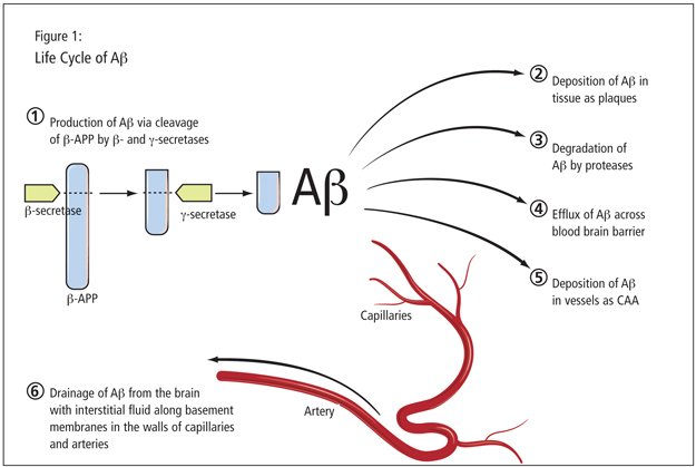
In cerebral amyloid angiopathy there is deposition of fibrillar peptides such as A, cystatin C, or transthyretin in the basement membranes present in the walls of capillaries and surrounding smooth muscle cells of arteries.26 The pattern of deposition of fibrillar amyloid peptides mirrors exactly the drainage pathways outlined by tracers after their injection into the brain parenchyma in experimental studies this suggests that CAA reflects a failure of clearance of proteins along the intramural periarterial drainage pathways.27 A recent study analyzing the changes that occur in the walls of arteries with CAA compared to normal ageing arteries demonstrated that Tissue Inhibitor of Metalloproteinase 3 is significantly elevated in CAA associated with Alzheimers disease and in Cerebral Autosomal-Dominant Arteriopathy with Subcortical Infarcts and Leukoencephalopathy , a condition in which peptides are also deposited in the vascular basement membranes.9,28,29 Recent studies demonstrate that in the Icelandic familial form of CAA, the deposition of the mutated cystatin C occurs specifically in the arterial basement membranes as well as basement membranes systemically, thus emphasizing the key role for extracellular matrix in the intramural periarterial drainage of proteins from the brain and in the pathogenesis of CAA.30
Kathryn L. Van Pelt, … Lisa M. Koehl, in, 2020
Also Check: What Color Ribbon Is Alzheimer’s
Cortical Microinfarcts And Other Vascular Lesions
Thirty-four cases presented with CMI of variable severity. In general, CMI were more numerous in cases with CAA than without , but without reaching statistical significance . Comparably, no significant differences were observed in the case of meningeal and intracerebral CAA . Finally, with the exception of a slight correlation between meningeal CAA and CMI in the frontal cortex , there was no association between the CAA and CMI in any of the other regions. Spearmans rho ranged from 0.12 to 0.134 for intracerebral CAA and from 0.12 to 0.134 for meningeal CAA in the other regions. Comparing different levels of severity , no statistical significance was revealed.
CMI in a case with severe CAA in the parietal cortex. Haematoxylin-eosin , and immunohistochemistry with anti-amyloid antibody 4G8 . Scale bar: : 500 µm : 200 µm.
There were only few vascular lesions of other types. Three cases showed microbleeds in the examined regions, and among them only 1 case presented with CAA. Among the 10 cases with macroscopic brain infarcts , 3 cases displayed CAA. Major intracerebral haemorrhages were found in 4 cases: 2 of them had both meningeal and intracerebral amyloid, one only meningeal amyloid, and the last one no amyloid deposition in vessels walls. The low number of these vascular lesions did not permit further statistical analyses.
The Impact Of Cerebral Amyloid Angiopathy In Various Neurodegenerative Dementia Syndromes: A Neuropathological Study
Jacques De Reuck
1Université de Lille 2, INSERM U1171, Degenerative & Vascular Cognitive Disorders, CHU Lille, 59000, France
Abstract
1. Introduction
The Boston criteria are used to suspect clinically the presence of cerebral amyloid angiopathy . It is diagnosed primarily as a cause of lobar cerebral haematoma in the elderly. Additionally cortical superficial siderosis , white matter changes , cortical microinfarcts , and cortical microbleeds are part of the pathological picture . However, on neuropathological examination of a large series of intracerebral haematomas only 9.7% are found to be due to CAA . In a small series of 13 patients with clinically suspected CAA the diagnosis could be confirmed by postmortem examination of the brain . Overall, the incidence of CAA in the elderly varies from 31.7% up to 78,9% according to different studies . In an older study the incidence of CAA in Alzheimers disease is observed in 25.6% . Also, CAA is found to be associated to 50% of brains with Lewy body disease and 25% of cases with progressive supranuclear palsy .
Whether CoMIs are significantly increased in Alzheimer patients with and without CAA is still a matter of debate .
No postmortem study has been performed about the impact of the degree of severity of CAA and its frequency in different neurodegenerative diseases.
2. Material and Methods
Main age and gender distribution was also compared between the different groups.
3. Results
| Items |
4. Discussion
Data Availability
You May Like: Alzheimers Awareness Ribbons
Uncommon Presentations Of Caa
Uncommon presentations of CAA include the following:
-
CAA can be associated with ischemic strokes in some of these patients, a coexistent vasculitis can be found the causal relationship with CAA is unclear
-
CAA is found in patients with autosomal dominant dementia, spasticity, and ataxia without ICH
-
CAA is reported in patients with vascular malformations, postirradiation necrosis, spongiform encephalopathies, and dementia pugilistica
-
CAA can present as a mass lesion, such as an amyloidoma with accumulation of amyloid in the brain parenchyma, or as edema and gliosis secondary to the vascular lesion
-
CAA can manifest as a reversible leukoencephalopathy, with rapid progression of symptoms and imaging abnormalities, followed by dramatic improvement
Symptoms Of Intracranial Hemorrhage
CAA most often comes to clinical attention because of ICH. Symptoms range from transient weakness to coma, depending on the size and location of the hemorrhage. Patients may have recurrent episodes.
The most common symptom at onset is headache , with the location of the pain varying in accordance with the location of the hematoma, as follows:
-
Frontal hematomas – Bifrontal headache pain
-
Parietal bleeds – Usually unilateral temple pain
-
Temporal hematomas – Ipsilateral eye and ear pain
-
Occipital bleeds – Ipsilateral eye pain
Vomiting tends to occur early. Seizures occur at onset in 16-36% of patients. Seizures are most commonly partial, with symptoms determined by the location of the ICH. As many as half of the patients present in status epilepticus.
You May Like: Alzheimer Awareness Ribbon Color
How Is Congophilic Amyloid Angiopathy Different From Other Diseases
The term congophilic is sometimes used because the presence of the abnormal aggregations of amyloid can be demonstrated by microscopic examination of brain tissue after staining with Congo red. The amyloid material is only found in the brain and as such the disease is not related to other forms of amyloidosis.
What Kind Of Angiopathy Is Amyloid Beta Peptide
Cerebral amyloid angiopathy. Cerebral amyloid angiopathy , is a form of angiopathy in which amyloid beta peptide deposits in the walls of small to medium blood vessels of the central nervous system and meninges.
The aim in cerebral amyloid angiopathy is to treat the symptoms, as there is no current cure. Physical, occupational and/or speech therapy may be helpful in the management of this condition.
Also Check: Mri Alzheimer’s Vs Normal
Is Neurodegeneration In Caa Solely A Function Of Concomitant Ad Pathology
Atrophy and cognitive impairment in CAA could result from either of three pathways, or a combination: a) the direct effects of tissue destruction by ICHs, b) the effects of concomitant AD pathology, or c) pathomechanisms other than ICH or AD .
Figure 3
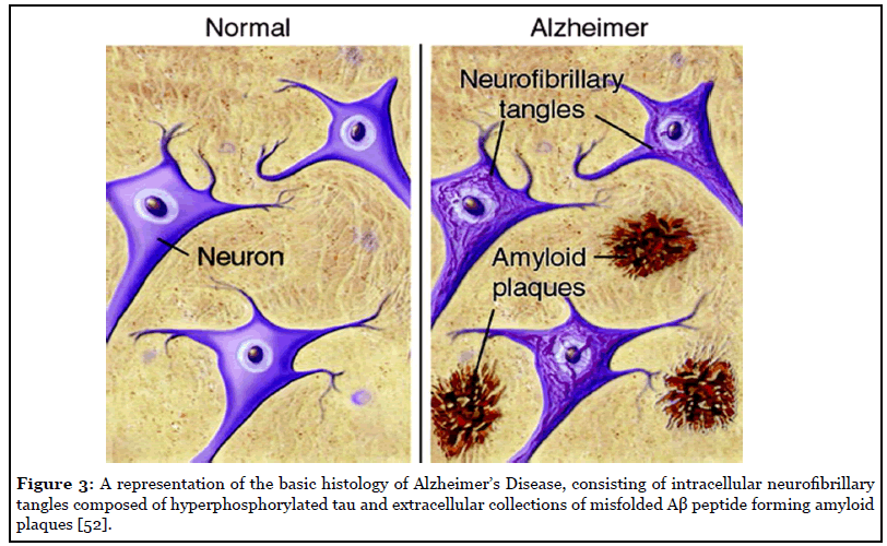
The potential contribution from AD pathology must be considered, because AD and CAA are sister diseases that share important aspects of their pathogenesis. Both are a consequence of cleavage of the amyloid precursor protein to form pathogenic Aβ which aggregates into β-amyloid. In the case of AD pathology, the β-amyloid aggregates in the brain parenchyma in the form of neuritic plaques. In the case of CAA, the β-amyloid aggregates in the walls of small arteries and arterioles. Most patients with CAA also have some degree of neuritic plaques, and most patients with AD have some degree of CAA .
However, there are also important contrasts between the two diseases . The beta-amyloid in CAA contains a higher proportion of Aβ1-40 compared to the beta-amyloid neuritic plaques . While the apolipoprotein E ε4 allele is a risk factor for both CAA and AD, the ε2 allele does not appear to protect against CAA and in fact may be associated with more severe vasculopathic changes .
| Cerebral amyloid angiopathy | |
|---|---|
| PET amyloid positive higher occipital : global ratio | PET amyloid positive lower occipital : global ratio |