Early Diagnosis Of Alzheimers Disease And Mild Cognitive Impairment
The typical reductions of hippocampal volume in MCI with an average Mini-Mental State Exam score of 25 are 10% to 15% and in AD with an average MMSE score of 20 are 20% to 25% . Measuring these significant reductions in the medial temporal lobe can be extremely useful for early diagnosis of AD and MCI. At present, diagnostic criteria for AD are based on the criteria in the Diagnostic and Statistical Manual of Mental Disorders, Fourth Edition , which are based primarily on clinical and psychometric assessment and do not use quantitative atrophy information available in sMRI scans. However, there is a proposal to add reliable biomarkers to the diagnostic criteria . One of the suggested features is the volume loss of medial temporal structures since measures of sMRI atrophy have accuracies of 70% to 90% in AD and 50% to 70% in amnestic MCI in distinguishing them from age-matched controls . All of the above-mentioned cross-sectional methods, except 3a, can be used as diagnostic metrics for AD and MCI.
Why Doctors Consider Mri To Detect Dementia
Medical experts will advise on the use of MRI when they suspect that a person has dementia.
MRI uses focused radio waves and magnetic fields to detect the presence of hydrogen atoms in tissues in the human body.
MRI scans also reveal the brains anatomic structure with 3D imaging allowing doctors to get a clear view of the current state of the organ.
This way, the doctor is able to rule out other health problems like hydrocephalus, hemorrhage, stroke, and tumors that can mimic dementia.
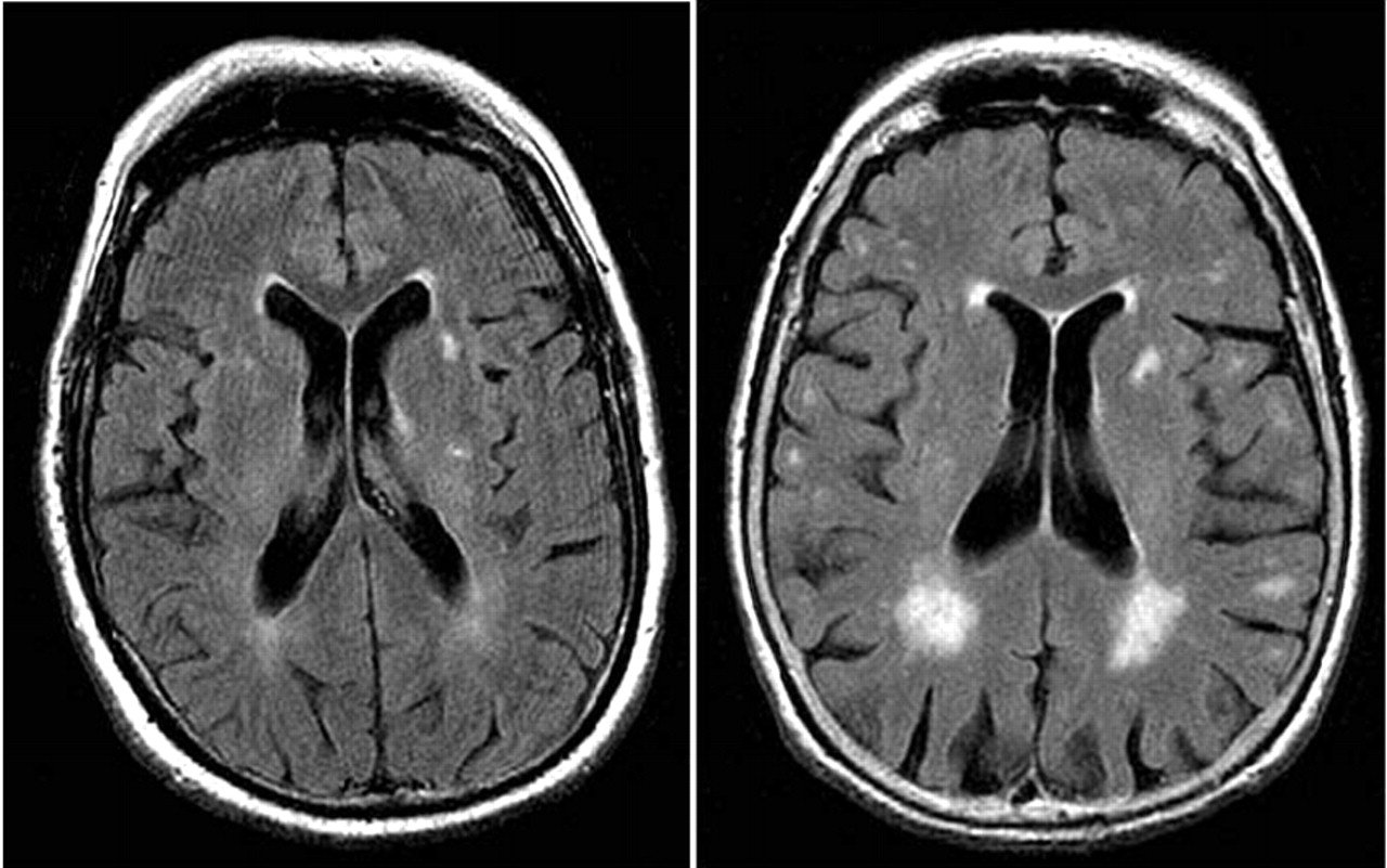
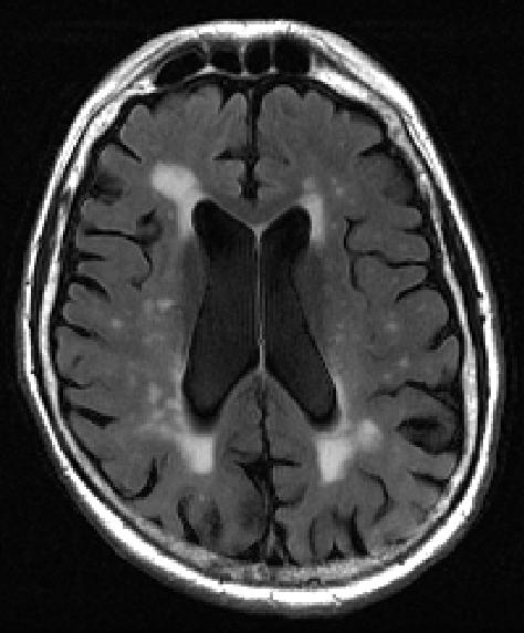
With these scans, physicians can also detect loss of brain mass that relates to different types of dementia.
fMRI records blood flow changes that are linked to the activities of the brain. This may help physicians differentiate dementia types.
Verywellhealth.com also suggests that MRI scans can at times identify reversible cognitive decline.
In such a case, a doctor will recommend appropriate treatment that will reverse this decline and restore cognitive functioning.
Treatments For Dementia With Lewy Bodies
Theres currently no cure for dementia with Lewy bodies or any treatment that will slow it down.
But there are treatments that can help control some of the symptoms, possibly for several years.
Treatments include:
- medicines to reduce hallucinations, confusion, drowsiness, movement problems and disturbed sleep
- therapies such as physiotherapy, occupational therapy and speech and language therapy for problems with movement, everyday tasks, and communication
Dont Miss: How To Move A Parent With Dementia To Assisted Living
Don’t Miss: Parkinsons And Alzheimers Together
Basics Of Fdg Pet As Applied To Ad
Brain FDG PET primarily indicates synaptic activity. Because the brain relies almost exclusively on glucose as its source of energy, the glucose analog FDG is a suitable indicator of brain metabolism and, when labeled with Fluorine-18 is conveniently detected with PET. The brains energy budget is overwhelmingly devoted to the maintenance of intrinsic, resting activity, which in cortex is largely maintained by glutamaturgic synaptic signaling . FDG uptake strongly correlates at autopsy with levels of the synaptic vesicle protein synaptophysin . Hence, FDG PET is widely accepted to be a valid biomarker of overall brain metabolism to which ionic gradient maintenance for synaptic activity is the principal contributor . In this context, a single, specific AD-related alteration in FDG metabolism has not been identified and therefore the FDG-PET abnormalities described below are assumed to be the net result of some combination of processes putatively involved in the pathogenesis of AD including, but not limited to, expression of specific genes, mitochondrial dysfunction, oxidative stress, deranged plasticity, excitotoxicity, glial activation and inflammation, synapse loss, and cell death.
You May Like: Did Margaret Thatcher Have Dementia
Brain Changes Evident In Scans Before Memory Cognitive Decline

- Date:
- Washington University School of Medicine
- Summary:
- Doctors may one day be able to gauge a patient’s risk of dementia with an MRI scan, according to a new study. Using a new technique for analyzing MRI data, researchers were able to predict who would experience cognitive decline with 89 percent accuracy.
One day, MRI brain scans may help predict whether older people will develop dementia, new research suggests.
In a small study, MRI brain scans predicted with 89 percent accuracy who would go on to develop dementia within three years, according to research at Washington University School of Medicine in St. Louis and the University of California San Francisco.
The findings, presented Sunday, Nov. 25 at the Radiological Society of North America meeting in Chicago, suggest that doctors may one day be able to use widely available tests to tell people their risk of developing dementia before symptoms arise.
“Right now it’s hard to say whether an older person with normal cognition or mild cognitive impairment is likely to develop dementia,” said lead author Cyrus A. Raji, MD, PhD, an assistant professor of radiology at Washington University’s Mallinckrodt Institute of Radiology. “We showed that a single MRI scan can predict dementia on average 2.6 years before memory loss is clinically detectable, which could help doctors advise and care for their patients.”
Story Source:
Cite This Page:
Also Check: Does Meredith Grey Have Alzheimer’s
Comparison Between Dlb And Pdd
Attempts to compare GM loss between DLB and PDD have revealed a pattern of more pronounced GM loss in DLB compared to PDD, which is in line with the fact that DLB encompasses greater amyloid burden . It is important to note, however, that localisations of GM reductions in DLB relative to PDD vary amongst different studies. For example, Burton et al. were unable to identify distinct cortical atrophy profiles of DLB and PDD , but Beyer et al. reported GM reductions in the temporal, parietal and occipital lobes in DLB using a voxel-based morphometry approach . Alongside the temporal and parietal atrophy, Lee et al. also reported occipital and striatal GM reductions in DLB . Studies investigating correlation patterns between brain structure and clinical and neuropsychiatric manifestations of the disease, revealed that decreased GM volume of the anterior cingulate, right hippocampus and amygdala were associated with cognitive performance , whilst reduced GM volume in the left precuneus and inferior frontal lobe correlated with visual hallucinations in DLB, but not in PDD .
You May Like: How Does Dementia Kill You
Experiment : Prediction Of Smci Vs Cad
The change in performances on the RAVLT-Im and ADAS13 tests are illustrated in Fig.;. Note the age-related decline in both the sMCI and the cAD subgroups, with the most severe impairments shown within the cAD group.
Figure; illustrates age-related tissue loss in the brain, with an almost linear shrinkage of the hippocampus volumes and a non-linear increase in the volume of the lateral ventricle . Overall, the most extensive losses are found among subjects in the cAD subgroup.
Inclusion of the cognitive trajectory features in the ensemble model gave \ for the accuracy, precision, recall and the \ scores. These scores changed to \, \, \ and \, respectively, when the longitudinal MRI features were added. The confusion matrices in Fig.; show a misclassification rate of \ for the subjects in both the cAD and the sMCI group when only the cognitive features were included, with a reduction to \ for the cAD subgroup and an increase to \ in the sMCI subgroup when the MRI features were added.
Figure 4
The trajectories for performances on the RAVLT-Im test and the ADAS13 test , with age at testing on the x-axis. The thick black curve is the cohort regression line, and thin grey lines are random effects for each subject. Severity of impairment is reflected by a lower score on the RAVLT test and a higher score on the ADAS13.
Also Check: What Is The Difference Between Dementia And Senility
Mri May Spot Early Signs Of Mental Decline
But scientists stress this doesn’t mean seniors should rush to get the scans, more study needed
HealthDay Reporter
TUESDAY, Oct. 7, 2014 — An MRI scan that measures blood flow in the brain may help predict which older adults are at risk for future memory loss, a preliminary study suggests.
The researchers found that, in some apparently healthy older adults, the MRI technique was able to pick up reductions in blood flow to a brain region linked to memory. And those people were more likely than their peers to show subtle memory loss 18 months later.
The results, reported online Oct. 7 in the journal Radiology, do not mean older adults should rush out to get brain scans, the researchers stressed.
But with more study, the MRI technique might prove useful for catching mental decline early. “That’s the aim in the long term,” said study leader Dr. Sven Haller, a senior physician at the University Hospitals of Geneva, in Switzerland.
For now, Haller said, the technology could be used in research — specifically, to select patients for clinical trials testing new drugs to stave off Alzheimer’s disease.
So far, Alzheimer’s drug trials have produced disappointing results, Haller pointed out.
“The problem is that one has to include patients at an early stage, because it is unlikely that a medication will recover already existing cognitive decline,” he said. “Yet it might — hopefully — decrease the speed of progression of cognitive decline,” he suggested.
How Is Alzheimers Disease Diagnosed And Evaluated
No single test can determine whether a person has Alzheimers disease. A diagnosis is made by determining the presence of certain symptoms and ruling out other causes of dementia. This involves a careful medical evaluation, including a thorough medical history, mental status testing, a physical and neurological exam, blood tests and brain imaging exams, including:
Dont Miss: Sandyside Senior Living
You May Like: Which Neurotransmitter Is Associated With Alzheimer’s
How Is Ftd Diagnosed
Assessments
Blood tests and a full physical examination are;important to rule out other possible causes of;symptoms.
A specialist normally an old age;psychiatrist or neurologist may think a person;has FTD after talking to them and to someone;who knows them well. The specialist will take a;detailed history of the persons symptoms and ask;questions to understand the persons behaviour;and abilities better.
Standard tests of mental abilities, which mostly;focus on memory loss, can be less helpful in;diagnosing FTD. More specialised tests of social;awareness or behaviour may be needed.
Scans
CT and MRI scans are used to see what;parts of the brain are most damaged. They can;also rule out other possible causes of a persons;symptoms, such as a stroke or tumour.If further tests are needed, more specialised brain;scans will be carried out, such as PET and SPECT to measure;the persons brain activity.
These scans are useful;as they may find lower activity in the frontal and/or;temporal lobes before a CT or MRI scan can find;changes to the brain tissue of these lobes.
Further;tests may include a lumbar puncture, which involves;collecting and examining liquid from inside the spine;and is carried out mainly in younger people.
Genetic testing
A specialist may recommend that a person with FTD;symptoms has a genetic test. This can show if the;persons condition is caused by a specific faulty;gene.
Post-mortem examination
Getting assessed for dementia
What Does Lewy Body Dementia Look Like
Lewy body dementia affects a persons ability to think and process information and it can negatively impact memory and alter personality. Though it shares aspects of other forms of dementia, there are distinct hallmarks of LBD. Lewy body dementia symptoms include:
- Fluctuating attention/alertness: These shifts can last hours or go on for days. The person may stare into space, appear lethargic or drowsy, and have hard-to-understand speech, appearing a lot like delirium. At other times, the person may have much more clarity of thought.
- Visual hallucinations: Often, these are very detailed hallucinations and visions of people or animals, and they can recur.
- Movement disorders: Parkinsons-like movement issues, such as muscle rigidity, tremors, falls, or a shuffling gait or way of walking, may occur.
Don’t Miss: Did Margaret Thatcher Have Dementia
What To Expect With A Head Ct Scan
If you decide to get a head CT scan, the process usually starts with contrast dye. Depending on your situation, this may be ingested orally or intravenously with a needle. This is simply dye that allows the images to show up on the screen.
Then, you get into the CT machine. While there are some standing CT machines, you generally need to use a machine that lets you lie down for a head scan. During the procedure, there is no pain, and in fact, all you have to do is stay still. Note that you may hear some noises, and some people feel anxious due to the confined space. If you anticipate feeling worried during the procedure, you may want to talk with your doctor about anti-anxiety medicine.
Facing the idea that you might have Alzheimers can be incredibly scary, but its important to remember that the earlier you detect the more likely you are to be able to manage the symptoms. To set up an appointment, contact American Health Imaging today.
Alternatives To A Head Scan

Arguably, a head CT scan is the most effective way to diagnose Alzheimers, but there are other options. There are a few cognitive tests that you can download, print, and take at home. Then, you bring these tests to your doctor to score. For instance, the SAGE test from the Ohio State University Wexner Medical Center is one option.
Unfortunately, an autopsy is the only way to conclusively diagnose Alzheimers disease. To get a quality diagnosis before death, you may need to combine several cognitive functioning tests. For example, a head CT test along with an assessment by your primary care doctor may be ideal.
Dont Miss: What Is The Color For Dementia
Also Check: What Is The Difference Between Dementia And Senility
Treatment And Other Helpful Guidelines
Medication
Mrs. Ws treatment team worked with her husband and her to preserve her independence, self-esteem, and quality of life as fully as possible. Although no medications carry a specific FDA indication or have been proven effective in reducing PCA symptoms, doctors often prescribe cholinesterase inhibitors such as donepezil , rivastigmine , or Razadyne or the glutamatergic medication memantine . This makes sense because AD plaques and tangles are so often the underlying cause of PCA.
Modifying the Home
As with AD, the clinical management of PCA goes far beyond the use of medication. A geriatric care manager in communication with Mrs. Ws treatment team helped to make her home a safer place for someone with her visual difficulties. Clutter was removed, and labels were applied to drawers so that she could find things more easily. Throw rugs were removed or replaced with non-skid floor coverings. Stickers were put on glass doors and large windows so that Mrs. W would see them more easily. ;Adequate lighting was arranged in all rooms with attention to reducing glare.
Other Helpful Tips
Developing Better And More Accurate Methods Of Diagnosis Is An Important Research Focus
Currently there is no single test that can accurately diagnose dementia.;
A detailed medical history, memory and thinking tests , laboratory tests and brain scans are typically used in the diagnosis process.
Current research into the diagnosis of Alzheimer’s disease and other types of dementia aims to develop better methods for accurate and earlier diagnosis. Early diagnosis of dementia is currently important to allow time for planning and to maximise the potential for treatment.
In the future, identification of individuals in the preclinical phase of dementia, before symptoms of cognitive decline are evident, will be possible. So we will be able to predict who is going to develop dementia, rather than wait to diagnose dementia after it emerges. This could lead to lifestyle prevention strategies in order to delay dementia onset, and to earlier use of therapies that slow or halt the disease process.
Also Check: Which Neurotransmitter Is Associated With Alzheimer’s
How A Head Ct Scan Can Detect Alzheimers Disease
A head CT scan looks at the structure of your brain. This scan can detect issues such as tumors, hemorrhages, and strokes, which can all mimic the symptoms of Alzheimers, but in addition to helping you rule out those conditions, a CT scan can also detect the loss of brain mass thats associated with Alzheimers disease.
What Is Magnetic Resonance Imaging
A magnetic resonance imaging scan is usually called an MRI. An MRI does not use radiation and is a noninvasive medical test or examination. The MRI machine uses a large magnet and a computer to take pictures of the inside of your body. Each picture or “slice” shows only a few layers of body tissue at a time. The pictures can then be examined on a computer monitor.
Pictures taken this way may help caregivers find and see problems in your body more easily. The scan usually takes between 15 to 90 minutes. Including the scan, the total examination time usually takes between 1.5 to 3 hours.
A substance called gadolinium is injected into a vein to help the physicians see the image more clearly. The gadolinium collects around cancer cells so they show up brighter in the picture. Sometimes a procedure called magnetic resonance spectroscopy is done during the MRI scan. An MRS is used to diagnose tumors based on their chemical make-up.
How does MRI work?
The MRI machine is a large, cylindrical machine that creates a strong magnetic field around the patient. This magnetic field, along with a radiofrequency, alters the hydrogen atoms’ natural alignment in the body.
A magnetic field is created and pulses of radio waves are sent from a scanner. The radio waves knock the nuclei of the atoms in the body out of their normal position; as the nuclei realign back into proper position, they send out radio signals.
Other related procedures that are used to assess the heart may include:
Recommended Reading: Smelling Farts Prevents Cancer
Can An Mri Detect Dementia
People who are suspected to have dementia will often ask can an MRI detect dementia.
This is because doctors often use brain scans to identify tumors, strokes, and other problems that might lead to dementia development.
MRI and CT scans are the most common types of brain scans that doctors use when they want to confirm whether a person has a neurodegenerative illness or not.