Brain Changes Evident In Scans Before Memory Cognitive Decline
- Date:
- Washington University School of Medicine
- Summary:
- Doctors may one day be able to gauge a patient’s risk of dementia with an MRI scan, according to a new study. Using a new technique for analyzing MRI data, researchers were able to predict who would experience cognitive decline with 89 percent accuracy.
One day, MRI brain scans may help predict whether older people will develop dementia, new research suggests.
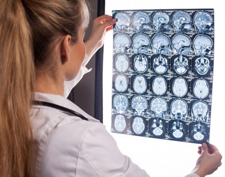
In a small study, MRI brain scans predicted with 89 percent accuracy who would go on to develop dementia within three years, according to research at Washington University School of Medicine in St. Louis and the University of California San Francisco.
The findings, presented Sunday, Nov. 25 at the Radiological Society of North America meeting in Chicago, suggest that doctors may one day be able to use widely available tests to tell people their risk of developing dementia before symptoms arise.
“Right now it’s hard to say whether an older person with normal cognition or mild cognitive impairment is likely to develop dementia,” said lead author Cyrus A. Raji, MD, PhD, an assistant professor of radiology at Washington University’s Mallinckrodt Institute of Radiology. “We showed that a single MRI scan can predict dementia on average 2.6 years before memory loss is clinically detectable, which could help doctors advise and care for their patients.”
Story Source:
Cite This Page:
Imaging In The Diagnosis And Prognosis Of Ad
The uncertainty inherent in a clinical diagnosis of AD has driven a search for diagnostic imaging markers. A definitive diagnosis still requires histopathological confirmation and the inaccessibility of the brain means imaging has a key role as a window on the brain. Historically, imagingfirst computed tomography and then MRIwas used only to exclude potentially surgically treatable causes of cognitive decline. Now its position in diagnosis also includes providing positive support for a clinical diagnosis of AD in symptomatic individuals by identifying characteristic patterns of structural and functional cerebral alterations. We can now also visualize the specific molecular pathology of the diseaseamyloid depositswith amyloid imaging. Alongside this increasing specificity for AD, imaging also contributes to differential diagnosis in practice by identifying alternative and/or contributory pathologies. Imaging is central to identifying vascular and non-AD degenerative pathologies and has helped in the recognition of the prevalence of mixed pathology in dementia.
Signs You May Need A Scan
If you have a family history of Alzheimers or dementia, you may want to get tested proactively so that you can determine if you have this condition. In other situations, you may want to get tested if you have any of the following symptoms:
- Memory loss
- Issues with depth perception
- Delusions or hallucinations
Many people believe that some memory loss is inevitable with aging, but this is simply not true. If you or your loved one is experiencing chronic or progressive memory loss or memory loss combined with the above symptoms, it may be a sign of Alzheimers.
Read Also: What Causes Alzheimer’s Disease In The Brain
Basics Of Structural Mri As Applied To Ad
MRI utilizes the fact that protons have angular momentum which is polarized in a magnetic field. This means that a pulse of radiofrequency can alter the energy state of protons and, when the pulse is turned off, the protons will, on returning to their energy stage, emit a radiofrequency signal. By a combination of different gradients and pulses, sequences can be designed to be sensitive to different tissue characteristics. In broad terms structural MRI in AD can be divided into assessing atrophy and changes in tissue characteristics which cause signal alterations on certain sequences such as white matter hyperintensities on T2-weighted MRI as a result of vascular damage. A number of MR sequences that are sensitive to microstructural change have shown alterations in AD. These sequences are already important research tools; however, they have not yet found a place in routine clinical practice in AD and they will not be considered further here.
Mri Brain Scans Accurate In Early Diagnosis Of Alzheimer’s Disease

Researchers advocate including imaging technology as diagnostic test
University of South Florida
image:;A normal MRI brain, showing no atrophy, depicts the three areas of interest in the brain’s medial temporal lobe: hippocampus ; entorhinal cortex and perirhinal cortex .view more;
Tampa, FL — MRI scans that detect shrinkage in specific regions of the mid-brain attacked by Alzheimer’s disease accurately diagnose the neurodegenerative disease, even before symptoms interfere with daily function, a study by the Florida Alzheimer’s Disease Research Center in Miami and Tampa found.
The study, reported earlier this month in the journal Neurology, adds to a growing body of evidence indicating MRI brain scans provide valuable diagnostic information about Alzheimer’s disease. The findings are important in light of many new disease-modifying drugs in trials — treatments that may prevent mild memory loss from advancing to full-blown dementia if administered early enough.
“This study demonstrates that MRI brain scans are accurate enough to be clinically useful, both in diagnosing Alzheimer’s disease itself at an early stage and in identifying people at risk of developing Alzheimer’s,” said Florida ADRC Center Director Huntington Potter, PhD, a neuroscientist at the Byrd Alzheimer’s Center and Research Institute, University of South Florida.
Journal
Neurology
Media Contact
Read Also: What Stage Of Alzheimer’s Is Aphasia
Schedule An Mri For Alzheimers Today
Early diagnosis is critical to slowing the progression of Alzheimers, and;an MRI of the head;is one of the best ways to do it. At Envision Imaging, were dedicated to providing world-class diagnostic imaging to enhance the quality of life for our patients.
No matter which of;our many locations;you visit, youll receive only the very best service from our staff of professionals who understand the stress that can surround a persons visit, so we ensure each client gets focused service with an excellent quality of care.
Find a;location near you;to schedule your MRI appointment today.
Alternatives To A Head Scan
Arguably, a head CT scan is the most effective way to diagnose Alzheimers, but there are other options. There are a few cognitive tests that you can download, print, and take at home. Then, you bring these tests to your doctor to score. For instance, the SAGE test from the Ohio State University Wexner Medical Center is one option.
Unfortunately, an autopsy is the only way to conclusively diagnose Alzheimers disease. To get a quality diagnosis before death, you may need to combine several cognitive functioning tests. For example, a head CT test along with an assessment by your primary care doctor may be ideal.
You May Like: How To Calm Down A Dementia Person
How A Computed Tomography Scan Can Help Diagnose Alzheimer’s
The next useful study that you can use in order to diagnose Alzheimer’s Disease would be a Computed Tomography scan, better known as the CT scan. This is an investigation that is more readily used in hospitals because of the speed and comfort for both the patient and the doctor. Unlike the MRI scan, this type of investigation does not require the use of a magnetic field, and therefore;patients with;metal implants of any type have no contraindications for this investigation.
A CT scan is an investigation that makes use of the simple X-ray but on a much larger scale. Patients will be passed through a scanner that will take pictures using multiple X-rays and the system will then actually build a picture of what the internal structures appear to be. This is a much quicker examination for patients and can be completed in as little as 15 minutes in most cases.;
What Did The Scientists Discover
Magnetic Resonance Imaging can be used to measure changes in brain microstructures of mice affected by Alzheimer’s disease, an inflammatory brain disease with effects that disrupt the limbic functions of emotion, learning, and memory.
THE TOOLS THEY USED
This research was conducted in the at the MagLab’s located at the University of Florida.
Read Also: Are Men Or Women More Likely To Get Alzheimers
How Mris Show Risk For Dementia
In the study, researchers analyzed MRI scans for physical signs that the patient is likely to experience cognitive decline. They did this using a technique called diffusion tensor imaging. This method assesses the movement of water molecules along the white matter tracts of the brain. If the molecules are not moving, it suggests that the person might have damage to the white tracts, which could mean cognition issues.
Significant damage to the white matter is a signal of cognitive decline. Using the Alzheimers Disease Neuroimaging Initiative, the study team identified ten people whose cognitive skills deteriorated over two years and matched them with ten other people whose skills were intact.
Analyzing the diffusion tensor MRI scans taken just before the two-year period, researchers found the significance of the change in white matter. When researchers detected more damage, they also observed greater cognitive decline. They repeated their analysis in a separate sample of 61 individuals and found that they could predict cognitive decline with 89 to 95 percent accuracy.
Raji and his team say what they need today is more research. Their priority is getting more control subjects and developing computerized tools that can reliably compare the scans to a baseline.
How Is Alzheimer’s Disease Diagnosed And Evaluated
No single test can determine whether a person has Alzheimer’s disease. A diagnosis is made by determining the presence of certain symptoms and ruling out other causes of dementia. This involves a careful medical evaluation, including a thorough medical history, mental status testing, a physical and neurological exam, blood tests and brain imaging exams, including:
Recommended Reading: What Is The 7th Stage Of Alzheimer’s
Utility Of Structural Mri In The Study Of Ad
Atrophy in AD
Progressive cerebral atrophy is a characteristic feature of neurodegeneration that can be visualized in life with MRI . The major contributors to atrophy are thought to be dendritic and neuronal losses. Studies of regional MRI volumes have shown these are closely related to neuronal counts at autopsy . The pattern of loss differs between diseases reflecting selective neuronal vulnerability and/or regional disease expression. AD is characterized by an insidious onset and inexorable progression of atrophy that is first manifest in the medial temporal lobe . The entorhinal cortex is typically the earliest site of atrophy, closely followed by the hippocampus, amygdala, and parahippocampus . Other structures within the limbic lobe such as the posterior cingulate are also affected early on. These losses then spread to involve the temporal neocortex and then all neocortical association areas usually in a symmetrical fashion. This sequence of progression of atrophy on MRI most closely fits histopathological studies that have derived stages for the spread of neurofibrillary tangles . Nonetheless, a significant minority of AD cases have atypical presentations and in these cases the pattern of atrophy accords with clinical phenotype: with language presentations particularly having left temporal atrophy and visual variants having posterior cortical atrophy.
Measuring Progression in AD with Structural MRI
Availability and Utility of Structural MRI
Can An Mri Detect Alzheimers Disease

By Darlene Ortiz 8 am on February 16, 2015
Alzheimers disease , which ranks as the fourth most common cause of death in the United States, can be difficult to definitively diagnose. The disease, which is characterized by the progressive onset of dementia, causes an individual to exhibit a number of symptoms ranging from confusion and forgetfulness to changes in mood and behavior.
As a leading provider of Alzheimers care in Jefferson County, were always looking to share some of the new and innovative diagnosis and treatment options in regards to memory care. Today, were going share some insight into using MRIs to detect Alzheimers disease its early stages doing so can help to enhance future health and quality of life for seniors and aging adults.
There is no single method used to prove that an individual has Alzheimers. Although a complete assessment usually includes several techniques such as genetic testing, neurological screening and a mental status exam brain imaging techniques like MRI and CT are playing an increasingly important part in the medical communitys efforts to better understand and diagnose Alzheimers.
For more information about Alzheimers care or to learn more about caring for a senior adult with a memory condition, visit our website at www.homecareassistancejeffersonco.com or contact us directly at 303-987-5992 and schedule a complimentary, no-obligation consultation.
Don’t Miss: Do Alzheimer Patients Talk In Their Sleep
Measure Volume In The Brain
An MRI can provide the ability to view the brain with 3D imaging. It can measure the size and amount of cells in the hippocampus, an area of the brain that typically shows atrophy during the course of Alzheimer’s disease. The hippocampus is responsible for accessing memory which is often one of the first functions to noticeably decline in Alzheimer’s.;
An MRI of someone with Alzheimer’s disease may also show parietal atrophy. The parietal lobe of the brain is located in the upper back portion of the brain and is responsible for several different functions including visual perception, ordering and calculation, and the sense of our body’s location.
Basics Of Fdg Pet As Applied To Ad
Brain FDG PET primarily indicates synaptic activity. Because the brain relies almost exclusively on glucose as its source of energy, the glucose analog FDG is a suitable indicator of brain metabolism and, when labeled with Fluorine-18 is conveniently detected with PET. The brains energy budget is overwhelmingly devoted to the maintenance of intrinsic, resting activity, which in cortex is largely maintained by glutamaturgic synaptic signaling . FDG uptake strongly correlates at autopsy with levels of the synaptic vesicle protein synaptophysin . Hence, FDG PET is widely accepted to be a valid biomarker of overall brain metabolism to which ionic gradient maintenance for synaptic activity is the principal contributor . In this context, a single, specific AD-related alteration in FDG metabolism has not been identified and therefore the FDG-PET abnormalities described below are assumed to be the net result of some combination of processes putatively involved in the pathogenesis of AD including, but not limited to, expression of specific genes, mitochondrial dysfunction, oxidative stress, deranged plasticity, excitotoxicity, glial activation and inflammation, synapse loss, and cell death.
Don’t Miss: How To Deal With Someone With Dementia
Current Practice In Diagnosing Dementia
The remainder of this information will provide an overview of the diagnosis process and a guide to what happens after diagnosis.;
It is important to remember that there is no definitive test for diagnosing Alzheimers disease or any of the other common causes of dementia. Findings from a variety of sources and tests must be pooled before a diagnosis can be made, and the process can be complex and time consuming. Even then, uncertainty may still remain, and the diagnosis is often conveyed as possible or probable. Despite this uncertainty, a diagnosis is accurate around 90% of the time.;
People with significant memory loss without other symptoms of dementia, such as behaviour or personality changes, may be classified as having a Mild Cognitive Impairment . MCI is a relatively new concept and more research is needed to understand the relation between MCI and later development of dementia. However, MCI does not necessarily lead to dementia and regular monitoring of memory and thinking skills is recommended in individuals with this diagnosis.;;
What Are The Benefits Of An Early Alzheimer’s Diagnosis
Early, accurate diagnosis is beneficial for several reasons. Beginning treatment early in the disease process may help preserve daily functioning for some time, even though the underlying Alzheimers process cannot be stopped or reversed.
Having an early diagnosis helps people with Alzheimers and their families:
Also Check: Does Dementia Shorten Your Lifespan
Why Doctors Consider Mri To Detect Dementia
Medical experts will advise on the use of MRI when they suspect that a person has dementia.
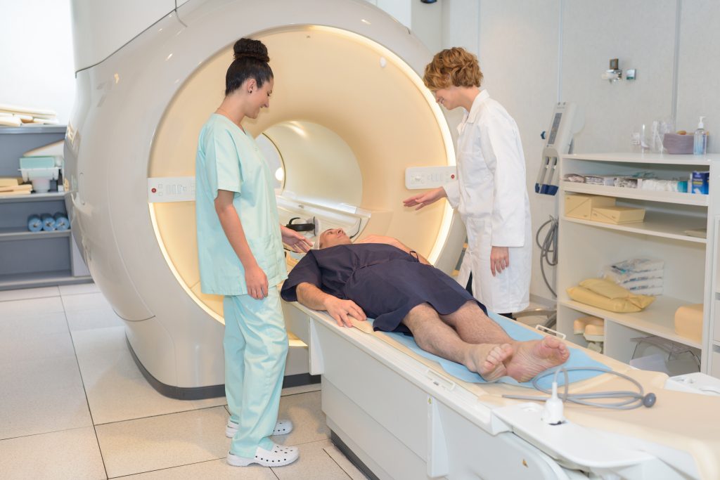
MRI uses focused radio waves and magnetic fields to detect the presence of hydrogen atoms in tissues in the human body.
MRI scans also reveal the brains anatomic structure with 3D imaging allowing doctors to get a clear view of the current state of the organ.
This way, the doctor is able to rule out other health problems like hydrocephalus, hemorrhage, stroke, and tumors that can mimic dementia.
With these scans, physicians can also detect loss of brain mass that relates to different types of dementia.
fMRI records blood flow changes that are linked to the activities of the brain. This may help physicians differentiate dementia types.
Verywellhealth.com also suggests that MRI scans can at times identify reversible cognitive decline.
In such a case, a doctor will recommend appropriate treatment that will reverse this decline and restore cognitive functioning.
What To Expect With A Head Ct Scan
If you decide to get a head CT scan, the process usually starts with contrast dye. Depending on your situation, this may be ingested orally or intravenously with a needle. This is simply dye that allows the images to show up on the screen.
Then, you get into the CT machine. While there are some standing CT machines, you generally need to use a machine that lets you lie down for a head scan. During the procedure, there is no pain, and in fact, all you have to do is stay still. Note that you may hear some noises, and some people feel anxious due to the confined space. If you anticipate feeling worried during the procedure, you may want to talk with your doctor about anti-anxiety medicine.
Facing the idea that you might have Alzheimers can be incredibly scary, but its important to remember that the earlier you detect the more likely you are to be able to manage the symptoms. To set up an appointment, contact American Health Imaging today.
You May Like: How Long Does Uti Dementia Last