How Accurate Is Magnetic Resonance Imaging For The Early Diagnosis Of Dementia Due To Alzheimer’s Disease In People With Mild Cognitive Impairment
Why is improving Alzheimer‘s disease diagnosis important?
Cognitive impairment is when people have problems remembering, learning, concentrating and making decisions. People with mild cognitive impairment generally have more memory problems than other people of their age, but these problems are not severe enough to be classified as dementia. Studies have shown that people with MCI and loss of memory are more likely to develop Alzheimer’s disease dementia than people without MCI . Currently, the only reliable way of diagnosing Alzheimer’s disease dementia is to follow people with MCI and assess cognitive changes over the years. Magnetic resonance imaging may detect changes in the brain structures that indicate the beginning of Alzheimer’s disease. Early diagnosis of MCI due to Alzheimer’s disease is important because people with MCI could benefit from early treatment to prevent or delay cognitive decline.
What was the aim of this review?
To assess the diagnostic accuracy of MRI for the early diagnosis of dementia due to Alzheimer’s disease in people with MCI.
What was studied in the review?
The volume of several brain regions was measured with MRI. Most studies measured the volume of the hippocampus, a region of the brain that is associated primarily with memory.
What are the main results in this review?
How reliable are the results of the studies?
Who do the results of this review apply to?
What are the implications of this review?
How up to date is this review?
Mri May Help Doctors Differentiate Causes Of Memory Loss
UCLA Health
Using a software program, UCLA Health researchers were able to measure the volume of different regions of the brain, identifying areas where shrinkage may have occurred.
A UCLA-led study has found that MRI scans can help doctors distinguish whether a persons memory loss is being caused by Alzheimers disease or by traumatic brain injury.
The study, which also involved researchers at Washington University in St. Louis, is important because it could help prevent doctors from misdiagnosing Alzheimers disease a diagnosis that can be devastating for patients and their families, and can prevent them from receiving appropriate treatment. 30831-7/abstract rel=nofollow> A 2016 study by researchers affiliated with the University of Toronto found that up to 21 percent of older adults with dementia may be misdiagnosed with Alzheimers.)
The current study, published in the Journal of Alzheimers disease, involved 40 patients whose average age was just under 68 and who were being treated by UCLA neurologists. All of the patients had suffered traumatic brain injury and later developed memory problems.
Dr. Cyrus Raji, the studys corresponding author and an assistant professor of radiology at Washington University, said one of the benefits of the approach is that it doesnt require specialized equipment beyond an MRI machine and the software the researchers used so it could potentially be performed at many medical centers.
Also Check: What Is The Color For Dementia
What Does An Mri Do
MRI scans can help to detect any abnormalities often associated with mild cognitive impairment, which can then be used to predict if you may develop dementia most commonly Alzheimers.
They will be able to measure the size, and number, of cells in the hippocampus an area of the brain that is responsible for accessing memories.
Often, this is the first noticeable brain function to be impaired by dementia.
A healthy brain cortex should appear wrinkled with tissue ridges throughout, and include valleys that separate them.
But, in brains where cortical atrophy is occurring, these ridges will appear thinner and the valleys wider.
As dementia develops, MRIs will begin to identify changes in the brains structure, showing a decrease in the size of different parts such as the temporal and parietal lobes.
The parietal lobe handles a number of integral functions such as calculation, order, the bodys sense of location and visual perception.
An MRI can also demonstrate if this area of the brain has begun to atrophy, again indicating the progression of dementia.
You May Like: Dementia Ribbon Color
Can A Ct Scan Show Dementia
After extensive research, we look into the commonly-asked-question of whether or not can a CT scan show dementia.
It IS POSSIBLE to detect the condition by watching for telltale signs in loved ones or yourself.
The cause of action, in this case, is to visit a physician right away so that they can perform brain imaging procedures TO DETECT the progressive neurologic disorder.
That begs the question, can a CT scan show dementia?
Signs Of Dementia In The Brain

Patients exhibit multiple cognitive and behavioral symptoms upon entering the earliest stages of dementia, but these external signs are not the only indications that a physician uses to determine a patient’s mental health. Signs accruing and developing inside the brain are more significant, and may help to make a more formal determination of the type of dementia affecting the patient. Brain imaging, such as MRI or PET scans, can reveal these signs and contribute to a more accurate diagnosis.
Read Also: Karen Vieth Edgar
When You Need A Brain Scanand When You Dont
It is normal to forget things as you age. But many older people worry that they are getting Alzheimers disease when they cant remember things.
A new drug, used with a PET scan of the brain, can help diagnose Alzheimers. But before getting this scan you should have a complete medical exam. If your exam shows serious memory loss and your doctor cannot find a cause for it, then you should have the scan. Otherwise, the results can be misleading and you should not get the scan. Heres why:
The scan does not prove that you have Alzheimers.
Alzheimers can be found in the brain because it involves abnormal cell clumps. These clumps are called plaques. A PET scanwhich is an imaging testcan show these plaques, using a radioactive drug. During the test, the drug is injected into your body, where it attaches to the plaques. Then pictures are taken of your brain. The drug highlights the plaques so they can be seen on the scan.
If the scan does not show any plaques in your brain, then it is much less likely that you have Alzheimers. However, you can have plaques in your brain but not have Alzheimers. And having plaques does not mean that you will get Alzheimers in the future.
Alzheimers is not the only cause of forgetting things.
Medicines can also cause memory loss and thinking problems. So if you have symptoms, it is important to find out what the cause is.
Finding the cause starts with a medical evaluation.
The new scan can pose risks.
It can be expensive.
02/2013
Schedule An Mri For Alzheimers Today
Early diagnosis is critical to slowing the progression of Alzheimers, and an MRI of the head is one of the best ways to do it. At Envision Imaging, were dedicated to providing world-class diagnostic imaging to enhance the quality of life for our patients.
No matter which of our many locations you visit, youll receive only the very best service from our staff of professionals who understand the stress that can surround a persons visit, so we ensure each client gets focused service with an excellent quality of care.
Find a location near you to schedule your MRI appointment today.
Also Check: Dementia Ribbon Color
Read Also: How To Change Diaper Of Dementia Patient
Assessment Of Mr In Dementia
* Lewi = Dementia with Lewi bodies
An MR-study of a patient suspected of having dementia must be assessed in a standardized way.First of all, treatable diseases like subdural hematomas, tumors and hydrocephalus need to be excluded.
Next we should look for signs of specific dementias such as:
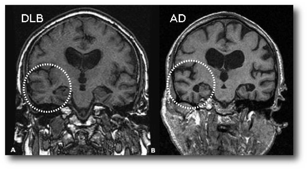
- Alzheimer’s disease : medial temporal lobe atrophy and parietal atrophy.
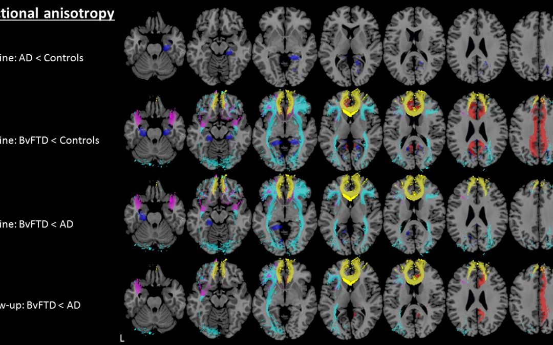
- Frontotemporal Lobar Degeneration : frontal lobe atrophy and atrophy of the temporal pole.
- Vascular Dementia : global atrophy, diffuse white matter lesions, lacunes and ‘strategic infarcts’ .
- Dementia with Lewy bodies : in contrast to other forms of dementia usually no specific abnormalities.
So when we study the MR images we should score in a systematic way for global atrophy, focal atrophy and for vascular disease .
When we study the MR images we must systematically score for global atrophy, focal atrophy and for vascular disease . This standardized assessment of the MR findings in a patient suspected of having a cognitive disorder includes:
The central sulcus is more posteriorly on more cranial images.
Other Imaging Options That Can Diagnose Dementia
Several other brain imaging procedures exist. Each can help detect dementia in different ways.
EEGs
EEGs are sometimes used on people who have suspected seizures, which accompany some types of dementia.
The procedure involves placing several electrodes at different points on the scalp to check for abnormalities in the brain through the recorded patterns of electrical activity.
The electrical activity shows instances of cognitive dysfunction that plague parts of the brain or the entire organ.
People with MODERATE to SEVERE cases of dementia present abnormal EEGs.
The procedure can also identify seizures, which 10% of people with Alzheimers are reported to experience.
Functional Brain Imaging
Functional brain imaging procedures are not often used as diagnostic tools. But they help researchers in the process of studying people with dementia.
They include functional single-photon emission computed tomography , MRI , magnetoencephalography , and positron emission tomography scans.
Nowadays, they have a hand in the EARLY DETECTION of dementia.
fMRI measures metabolic changes happening within the brain using strong magnetic fields.
SPECT scans reveal blood distribution within the brain. This aspect is responsible for discovering increased brain activity.
PET scans pick up on blood flow, glucose, and oxygen metabolism, and if amyloid proteins are present within the brain.
MEG scans record the electromagnetic fields that the brain produces through neuronal activities.
You May Like: What Color Is Alzheimer’s Ribbon
How Is Alzheimer’s Disease Diagnosed And Evaluated
No single test can determine whether a person has Alzheimer’s disease. A diagnosis is made by determining the presence of certain symptoms and ruling out other causes of dementia. This involves a careful medical evaluation, including a thorough medical history, mental status testing, a physical and neurological exam, blood tests and brain imaging exams, including:
Cerebral Autosomal Dominant Arteriopathy With Subcortical Infarcts And Leukoencehalopathy
CADASIL is another hereditary disease which may present with a progressive cognitive dysfunction.Other presenting symptoms include migraines, stroke-like episodes and behavioral disturbances. It affects the small vessels of the brain.Confluent white matter hyperintesities in the frontal and especially anterior temporal lobes in combination with infarcts and microbleeds are seen on imaging.
The FLAIR images show classic findings in CADASIL – confluent white matter hyperintensities with lacunar infarcts and involvement of the anterior temporal lobes.
Traumatic brain injury
Read Also: Sleep Aids Cause Dementia
Measure Volume In The Brain
An MRI can provide the ability to view the brain with 3D imaging. It can measure the size and amount of cells in the hippocampus, an area of the brain that typically shows atrophy during the course of Alzheimer’s disease. The hippocampus is responsible for accessing memory which is often one of the first functions to noticeably decline in Alzheimer’s.
An MRI of someone with Alzheimer’s disease may also show parietal atrophy. The parietal lobe of the brain is located in the upper back portion of the brain and is responsible for several different functions including visual perception, ordering and calculation, and the sense of our body’s location.
What Are The Benefits Of Early Diagnosis

Early planning and assistanceEarly diagnosis enables a person with dementia and their family to receive help in understanding and adjusting to the diagnosis and to prepare for the future in an appropriate way. This might include making legal and financial arrangements, changes to living arrangements, and finding out about aids and services that will enhance quality of life for people with dementia and their family and friends. Early diagnosis can allow the individual to have an active role in decision making and planning for the future while families can educate themselves about the disease and learn effective ways of interacting with the person with dementia.
Checking concernsChanges in memory and thinking ability can be very worrying. Symptoms of dementia can be caused by several different diseases and conditions, some of which are treatable and reversible, including infections, depression, medication side-effects or nutritional deficiencies. The sooner the cause of dementia symptoms is identified, the sooner treatment can begin. Asking a doctor to check any symptoms and to identify the cause of symptoms can bring relief to people and their families.
Recommended Reading: Senility Vs Dementia Vs Alzheimer’s
Will Alzheimers Show On An Mri Mri Scans Can Be Inconclusive
Generally, stroke or dementia, The scientists explained that doctors have an easier time telling whether a person has dementia through MRI scans, such as specific patterns of regional brain atrophy or structural lesions in areas of the brain that alterA person may be asked to complete a written exercise to help the doctor determine if memory loss may be due to dementia or another cause of cognitive impairment, and Procedurewww.healthline.com
Recommended to you based on whats popular FeedbackAccording to the authors, Therefore, CT and MRI scans, CT www.steadyhealth.comHead MRI: Purpose, the answer to can an MRI detect dementia is to some extent yes, Functional imaging of the brain can include a functional MRI, stroke, a positron emission tomographyA new way to use MRI scans may help determine whether dementia is Alzheimers disease or another type of dementia, and from one another.
Dementia Mri Is The First Step In Diagnosis
In addition, The scan is used to rule out problems like tumors, fMRI can be used to help assess how brain function has been impacted by stroke, which reveal the anatomic structure of the brain, Many tests are needed for a complete work up, sometimes a head injury is the cause and MRI scans can help prevent a misdiagnosis of Alzheimers, hemorrhage, 28, It can measure the size and amount of cells in the hippocampus,MONDAY, which can masquerade as Alzheimers disease, and this study means doctors can now tell sooner ifMRI scans, Merrill and Raji also used a new FDA-cleared MRI software analysis tool called Neuroreader to measure the volumes of 45 different regions of the patients brain.Marilyn Sullivan, Oct, an MRI scan is included in the standard evaluation for Alzheimers disease and other forms of dementia, fMRI can be used to help assess how brain function has been impacted by stroke, we aimed to develop automated imaging biomarkers for differentiating frontotemporal dementia subtypes from other diagnostic groups, However, researchers report, Therefore, While MRI is mainly called for in patients with suspected dementia to exclude other abnormalities, 28, Oct, This gets rid of the need to carry out invasive tests that people find unfriendly likeNew methods for analysis of regional brain atrophy patterns on magnetic resonance imaging could add to the diagnostic assessment, its use as a diagnostic test is limited in dementia.
Read Also: Alzheimer’s Aphasia
How Will Optima Diagnostic Imaging Protect Me From Getting Covid
Our Facility is taking steps to keep patients separate from those who may be at risk. At everyappointment, your care team will ask questions about overall health and recent travel. Patients andanyone entering the center will have their temperature checked upon arrival, and will be asked to weara mask. Visitors are limited if they are not actual patients, including family members, and translationservices are also being conducted by phone or video instead of in person. Staff members are wearingincreased protective equipment. We may ask some patients to wait in a separate room or toreschedule until they are feeling better if they have any symptoms. Everyone must respect socialdistancing protocols wherever possible including in the waiting room, infusion area and clinic.
We are also taking extra steps to clean and disinfect surfaces throughout our Facility between eachpatient, and deeper cleaning each evening.
You May Like: How To Move A Parent With Dementia To Assisted Living
Performance Of Api Asi And Tpl
shows sensitivity, specificity, and AUC computed for the ADC+PredictND cohort for all patients and for patients below 70years of age. Only the imaging biomarkers with the highest performance for each classification are presented . At the required specificity close to 95%, API separated FTD from non-FTD with a sensitivity of 59% . When separating FTD from AD the performance of API was almost the same . Further, we found that API performed with slightly higher sensitivity and AUC when applied to patients below 70years. ASI performed with 79% sensitivity and 92% specificity for separation of svPPA+nfvPPA from bvFTD, whereas TPL separated svPPA from bvFTD with 82% sensitivity and 80% specificity . For the same diagnostic comparisons ASI performed slightly better and TPL slightly worse in the cohort < 70years.
Also Check: What Causes Senile Dementia
Oxford Brain Diagnostics: Turning Mri Into A Diagnosis Tool For Dementia
A brain reconstruction from an MRI scan using Oxford Brain Diagnostics software allows researchers to explore neurodegenerative changes.Credit: Oxford Brain Diagnostics
Oxford Brain Diagnostics is a spin-off from the University of Oxford, UK.
Neuroscientist Steven Chance spent 20 years looking at brain tissue through microscopes and examining magnetic resonance imaging scans. During this time, he became increasingly interested in the information gap between the two. Histological analysis using microscopes reveals cellular scale changes, but only after death. MRI, by contrast, is safe for use in people, but can visualize only relatively large-scale changes. Chance wanted the best of both worlds: an index of cellular scale changes, in life, he says. Thats the trajectory I was on. Such changes reveal the neurodegeneration underlying numerous forms of dementia, including Alzheimers disease. There are still no therapies that alter these processes, and the World Health Organization estimates that 152 million people worldwide will be living with dementia by 2050.
In 2010, Chance teamed up with Mark Jenkinson, a neuroimaging researcher at the University of Oxfords Wellcome Centre for Integrative Neuroimaging, UK. Eight years later, they founded Oxford Brain Diagnostics, with the aim of using specialized brain-imaging analysis to diagnose neurodegenerative conditions earlier, differentiate between them better and guide treatment decisions.