Perfusion Imaging And Amyloid Related Vascular Changes
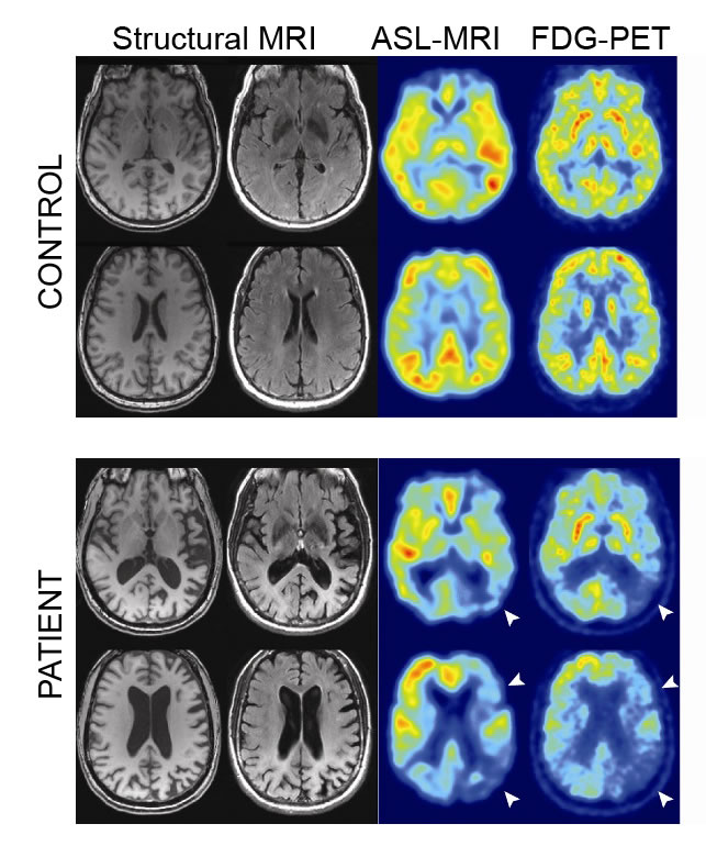
An emerging picture in AD research is the role of cerebral perfusion, with vascular changes occurring early in disease progression . In particular, global and regional cerebral hypoperfusion are associated with the disease, though debate continues as to whether this is a cause or effect . Either way, the observation that AD is thereby linked to modifiable vascular risk factors has attracted considerable attention. Hypoperfusion as measured by ASL has been consistently observed in the posterior cingulate, precuneus, inferior parietal, and lateral prefontal cortices . ASL findings compare well to FDG-PET , with recent data also suggesting the capacity of ASL to add to diagnostic power, with distinct patterns of perfusion in different dementias .
As well as atherosclerotic changes in people with AD, blood vessels can be affected by cerebral amyloid angiopathy or damage from amyloid-modifying therapies . Such changes include vasogenic oedema and microhaemorrhages .
With Alzheimers Disease Prevention Is Key
There are many ways to diagnose Alzheimers, although a definitive diagnosis cannot be achieved except in an autopsy, where brain tissue can be precisely examined. Typically, doctors use various tests to rule out conditions that could explain dementia-like symptoms.
Patients diagnosed with Alzheimers disease live another 4-8 years, on average, after their diagnosis.
However, you dont have to sit back and wait for Alzheimers to take its toll. Some patients live up to 20 years after their diagnosis!
Because most if not all conventional dementia treatments have about a 1% chance of success, the need for a revolutionary new approach is critical for treating dementia and Alzheimers.
At PrimeHealth, Dr. David Ward hosts an Alzheimers Prevention Program incorporating Dr. Dale Bredesens revolutionary, evidence-based lifestyle approach to optimize cognitive function. Schedule an appointment with us and learn more about PrimeHealths Prevention Program.
Reviewing Health History And Mental State
If you are experiencing troubling changes in memory, behavior, or thinking abilities, make an appointment to discuss your concerns with a doctor, even if it seems scary.
Primary-care physicians often oversee the diagnostic process but may refer patients to a specialist such as a neurologist, neuropsychologist, geriatrician, or geriatric psychiatrist.
An Alzheimers assessment will include the following:
- Medical and family history Your history includes past and present illnesses, any medications you take, and any conditions affecting other members of your family.
- Mood assessment The doctor will look for signs of depression or other mental-health disorders that can cause memory problems or other dementia-like symptoms.
Read Also: What Color Represents Dementia
Mri And Neuropsychological Data
For each subject of Table 1, and for each time point , structural MR images were downloaded from the ADNI data repository. According to the ADNI acquisition protocol , examinations were performed at 1.5 T using a T1-weighted sequence. We considered MR images that had undergone the following preprocessing steps: 3D gradwarp correction for geometry correction caused by gradient non-linearity , and B1 non-uniformity correction for intensity correction caused by non-uniformity . These preprocessing steps help improving the standardization among MR images from different MR sites and different platforms. MR images were downloaded in 3D NIfTI format. A further processing procedure was then performed on the downloaded images, this procedure consisting in: image re-orientation cropping skull-stripping image normalization to the MNI standard space by means of co-registration to the MNI template . MR images were then segmented into Gray Matter and White Matter tissue probability maps, and smoothed using an isotropic Gaussian kernel with Full Width at Half Maximum ranging from 2 to 12 mm3, with a step of 2 mm3. After this phase, all MR images resulted to be of size 121 × 145 × 121 voxels. The whole process was performed using the VMB8 software package installed on the Matlab platform . MRI volumes were visually inspected for checking homogeneity and absence of artifacts both before and after the pre-processing step.
The Limitations Of Amyloid Pet In Ad

Major deterrents to the widespread use of amyloid PET remain cost and availability. Availability has been improved by the development of F-18-labeled agents that can be distributed to PET scanners not associated with a cyclotron. Cost remains an issue, especially where CSF measurement of A42 can provide very similar information when the question is simply the presence or absence of brain A deposition. Being an early event in the pathogenesis of AD, amyloid PET is not a good surrogate marker of progression during the clinical stage of the disease . This role is filled much better by structural MRI and FDG PET . Similarly, amyloid imaging gives much more of a binary diagnostic readout than techniques such as MRI and FDG PET. That is, amyloid imaging has a certain specificity for the pathology of AD, but when that pathology is absent, a negative amyloid PET scan will be identical regardless of the non-AD etiology of the dementia. In contrast, MRI and FDG PET may give an indication of a frontotemporal or vascular pathology when an amyloid PET scan would be ambiguously negative in both cases. The threshold of sensitivity of amyloid PET has yet to be precisely determined, but it is clear that some level of amyloid deposition is histologically detectable prior to the in vivo signal becoming positive .
Read Also: Ribbon Color For Alzheimer’s
Can The Mindcrowd Test Be Used To Figure Out If A Medical Consultation Is Needed
No. This is not a diagnostic test. MindCrowd is a research test, and its not designed to diagnose Alzheimers.
Dont be afraid to take it, even if you find it difficult, because it doesnt mean you have dementia or even mild cognitive decline. But you are helping scientists learn more about how the brain ages.
Alzheimers diagnostic testing looks at many aspects of how your brain works and the MindCrowd test is only looking at two particular areas: Attention and Memory.
Mri Scans As Accurate As Lumbar Punctures In Identifying Alzheimers Or Ftld
They found that by studying the structural brain patterns the density of gray matter on the MRI scans, their predictions were 75% accurate when confirming diagnosis with people who had pathology-confirmed diagnoses and those with biomarker levels retrieved from lumbar punctures this shows that the new MRI use is as accurate as lumbar puncture methods.
McMillan said:
Developing a new method for diagnosis is important because potential treatments target the underlying abnormal proteins, so we need to know which disease to treat. This could be used as a screening method and any borderline cases could follow up with the lumbar puncture or PET scan.
This method would also be helpful in clinical trials where it may be important to monitor these biomarkers repeatedly over time to determine whether a treatment was working, and it would be much less invasive than repeated lumbar punctures.
Recommended Reading: What Color Ribbon Is Alzheimer’s
Future Directions In Diagnosis Research
Considerable research effort is being put into the development of better tools for accurate and early diagnosis. Research continues to provide new insights that in the future may promote early detection and improved diagnosis of dementia, including:
- Better dementia assessment tests that are suitable for people from diverse educational, social, linguistic and cultural backgrounds.
- New computerised cognitive assessment tests which can improve the delivery of the test and simplify responses.
- Improved screening tools to allow dementia to be more effectively identified and diagnosed by GPs.
- The development of blood and spinal fluid tests to measure Alzheimers related protein levels and determine the risk of Alzheimers disease.
- The use of sophisticated brain imaging techniques and newly developed dyes to directly view abnormal Alzheimers protein deposits in the brain, yielding specific tests for Alzheimers disease.
Signs You May Need A Scan
If you have a family history of Alzheimers or dementia, you may want to get tested proactively so that you can determine if you have this condition. In other situations, you may want to get tested if you have any of the following symptoms:
- Memory loss
- Issues with depth perception
- Delusions or hallucinations
Many people believe that some memory loss is inevitable with aging, but this is simply not true. If you or your loved one is experiencing chronic or progressive memory loss or memory loss combined with the above symptoms, it may be a sign of Alzheimers.
Don’t Miss: Shampoos Cause Alzheimer’s
How Is Alzheimer’s Disease Diagnosed
Doctors use several methods and tools to help determine whether a person who is having memory problems has possible Alzheimers dementia , probable Alzheimers dementia , or some other problem.
To diagnose Alzheimers, doctors may:
- Ask the person and a family member or friend questions about overall health, use of prescription and over-the-counter medicines, diet, past medical problems, ability to carry out daily activities, and changes in behavior and personality
- Conduct tests of memory, problem solving, attention, counting, and language
- Carry out standard medical tests, such as blood and urine tests, to identify other possible causes of the problem
- Perform brain scans, such as computed tomography , magnetic resonance imaging , or positron emission tomography , to rule out other possible causes for symptoms
These tests may be repeated to give doctors information about how the persons memory and other cognitive functions are changing over time. They can also help diagnose other causes of memory problems, such as stroke, tumor, Parkinsons disease, sleep disturbances, side effects of medication, an infection, mild cognitive impairment, or a non-Alzheimers dementia, including vascular dementia. Some of these conditions may be treatable and possibly reversible.
People with memory problems should return to the doctor every 6 to 12 months.
Promoting Early Diagnosis Of Dementia
The early symptoms of dementia can include memory problems, difficulties in word finding and thinking processes, changes in personality or behaviour, a lack of initiative or changes in day to day function at home, at work or in taking care of oneself. This information does not include details about all of these warning signs, so it is recommended that you seek other sources of information. If you notice signs in yourself or in a family member or friend, it is important to seek medical help to determine the cause and significance of these symptoms.
Obtaining a diagnosis of dementia can be a difficult, lengthy and intensive process. While circumstances differ from person to person, Dementia Australia believes that everyone has the right to:
- A thorough and prompt assessment by medical professionals,
- Sensitive communication of a diagnosis with appropriate explanation of symptoms and prognosis,
- Sufficient information to make choices about the future,
- Maximal involvement in the decision making process,
- Ongoing maintenance and management, and
- Access to support and services.
Read Also: Do Sleeping Pills Cause Dementia
Ai Algorithm Can Accurately Predict Risk Diagnose Alzheimers Disease
Researchers have developed a computer algorithm based on Artificial Intelligence that can accurately predict the risk for and diagnose Alzheimers disease using a combination of brain magnetic resonance imaging , testing to measure cognitive impairment, along with data on age and gender.
The AI strategy, based on a deep learning algorithm, is a type of machine learning framework. Machine learning is an AI application that enables a computer to learn from data and improve from experience. Alzheimers disease is the primary cause of dementia worldwide. One in 10 people age 65 and older has Alzheimers dementia. It is the sixth-leading cause of death in the United States.
If computers can accurately detect debilitating conditions such as Alzheimers disease using readily available data such as a brain MRI scan, then such technologies have a wide-reaching potential, especially in resource-limited settings, explained corresponding author Vijaya B. Kolachalama, PhD, assistant professor of medicine. Not only can we accurately predict the risk of Alzheimers disease but this algorithm can generate interpretable and intuitive visualizations of individual Alzheimers disease risk en route to accurate diagnosis, said Dr. Kolachalama.
The researchers believe their methodology can be extended to other organs in the body and develop predictive models to diagnose other degenerative diseases.
These findings appear online in the journal Brain.
Is Alzheimers Diagnosed With A Blood Test

The other biomarker test, uses either a blood sample or, more commonly, a cerebral spinal fluid sample. The CSF sample is obtained via a spinal tap.
We look for markers of Alzheimers disease in the blood and the spinal fluid samples. These are pieces of the plaques and tangles that might be circulating in the blood or spinal fluid.
Therefore, the patient can have a thinking and memory test, and then typically the physician will add on additional tests. One could be a PET scan of the brain and the other one could be a test of either the blood or cerebral spinal fluid.
Diagnosing Alzheimers disease is a process. And its typically a combination of these three approaches: Cognitive Testing, PET biomarkers and fluid either blood or spinal biomarkers.
In all cases, personal results are compared to norms. The person is compared to other healthy people in the population that are approximately just like them. Neurologists look at the levels of all of the things that they are measuring to determine if they think its Alzheimers disease.
And thats how a diagnosis goes.
Don’t Miss: Does Neil Diamond Have Alzheimer’s
Measure Volume In The Brain
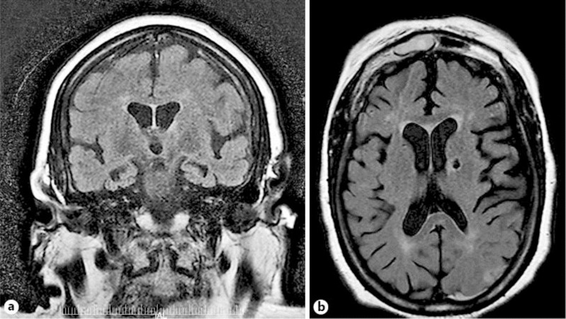
An MRI can provide the ability to view the brain with 3D imaging. It can measure the size and amount of cells in the hippocampus, an area of the brain that typically shows atrophy during the course of Alzheimer’s disease. The hippocampus is responsible for accessing memory which is often one of the first functions to noticeably decline in Alzheimer’s.
An MRI of someone with Alzheimer’s disease may also show parietal atrophy. The parietal lobe of the brain is located in the upper back portion of the brain and is responsible for several different functions including visual perception, ordering and calculation, and the sense of our body’s location.
What Is Alzheimer’s Disease
Alzheimer’s disease is the most common cause of dementia, a loss of brain function that affects memory, thinking, language, judgment and behavior. In Alzheimer’s disease, large numbers of neurons stop functioning, lose connections with other neurons, and die.
Irreversible and progressive, Alzheimer’s disease slowly destroys memory and thinking skills and, eventually, the ability to carry out the simplest tasks of daily living.
Although the cause of Alzheimer’s disease is unknown, scientists believe that a build-up of beta-amyloid plaques and neurofibrillary tangles in the brain are associated with the disease.
The stages of the disease typically progress from mild to moderate to severe. Symptoms usually develop slowly and gradually worsen over a number of years however, progression and symptoms vary from person to person. The first symptom of Alzheimer’s disease usually appears as forgetfulness.
Mild cognitive impairment is a stage between normal forgetfulness due to aging and the development of Alzheimer’s disease. People with MCI have mild problems with thinking and memory that do not interfere with everyday activities. Not everyone with MCI develops Alzheimer’s disease.
Other early symptoms of Alzheimer’s include language problems, difficulty performing tasks that require thought, personality changes and loss of social skills.
People with severe Alzheimer’s disease are unable to recognize family members or understand language.
Don’t Miss: Does Diet Coke Cause Alzheimer’s
How A Computed Tomography Scan Can Help Diagnose Alzheimer’s
The next useful study that you can use in order to diagnose Alzheimer’s Disease would be a Computed Tomography scan, better known as the CT scan. This is an investigation that is more readily used in hospitals because of the speed and comfort for both the patient and the doctor. Unlike the MRI scan, this type of investigation does not require the use of a magnetic field, and therefore patients with metal implants of any type have no contraindications for this investigation.
A CT scan is an investigation that makes use of the simple X-ray but on a much larger scale. Patients will be passed through a scanner that will take pictures using multiple X-rays and the system will then actually build a picture of what the internal structures appear to be. This is a much quicker examination for patients and can be completed in as little as 15 minutes in most cases.
How Is Alzheimers Disease Diagnosed
So little is known about Alzheimers that theres no single test to diagnose it. However, there are ways to test brain function, and these tests, along with examining symptoms and ruling out other potential conditions, are generally how physicians diagnose Alzheimers. Your family doctor can diagnose the disease, but psychologists, neurologists and geriatricians can also provide a diagnosis or a second opinion.
An examination for diagnosing Alzheimers disease can include:
- Complete and accurate medical history
- Test for mental status
- Blood tests
- Brain imaging
Brain imaging allows medical professionals to see how the brain is functioning and accurately spot any abnormalities. Alzheimers imaging can include:
Also Check: Does Bobby Knight Have Dementia
Basics Of Functional Mri As Applied To Ad
Functional MRI is being increasingly used to probe the functional integrity of brain networks supporting memory and other cognitive domains in aging and early AD. fMRI is a noninvasive imaging technique which provides an indirect measure of neuronal activity, inferred from measuring changes in blood oxygen leveldependent MR signal . Whereas fluoro-deoxy-d-glucose -PET is thought to be primarily a measure of synaptic activity, BOLD fMRI is considered to reflect the integrated synaptic activity of neurons via MRI signal changes because of changes in blood flow, blood volume, and the blood oxyhemoglobin/deoxyhemoglobin ratio . fMRI can be acquired during cognitive tasks, typically comparing one condition to a control condition , or during the resting state to investigate the functional connectivity within specific brain networks. Fc-MRI techniques examine the correlation between the intrinsic oscillations or time course of BOLD signal between brain regions , and have clearly documented the organization of the brain into multiple large-scale brain networks . Both task-related and resting fMRI techniques have the potential to detect early brain dysfunction related to AD, and to monitor therapeutic response over relatively short time periods however, the use of fMRI in aging, MCI, and AD populations thus far has been limited to a relatively small number of research groups.