The Role Of The Amygdala In Memory
Biological:Behavioural genetics · Evolutionary psychology · Neuroanatomy · Neurochemistry · Neuroendocrinology ·Neuroscience · Psychoneuroimmunology · Physiological Psychology · Psychopharmacology
The amygdala plays a key role in the modulation of memory consolidation. Following any learning event, the long-term memory for the event is not instantaneously formed. Rather, information regarding the event is slowly put into long-term storage over time, a process referred to as “memory consolidation”, until it reaches a relatively permanent state.
During the consolidation period, the memory can be modulated. In particular, it appears that emotional arousal following the learning event influences the strength of the subsequent memory for that event. Greater emotional arousal following a learning event enhances a person’s retention of that event. Experiments have shown that administration of stress hormones to a person immediately after the person learns something enhances that person’s retention when they are tested two weeks later.
Despite the importance of the amygdala in modulating memory consolidation, however, learning can occur without it, though such learning appears to be impaired.
Dementia And Emotional Responses
Dementia is a group of symptoms pointing to deterioration of parts of the brain that interferes with the way the person processes his or her environment. But while cognition and other functions may decline, the brains amygdala is often intact to the end of life. With the emotional center still open for business, the person experiences feelings. Some are positive , and others are negative .
Traditionally, the amygdala has been primarily associated with humans fight-or-flight response. It is an automatic, ingrained survival response to particular stimuli, and its something all humans experience. Under these current stresses related to the pandemic, some of us are irritable or angry and take these feelings out on others. Or, reactions fall under the flight category, manifesting in such ways as depression, lethargy, and social retreat.
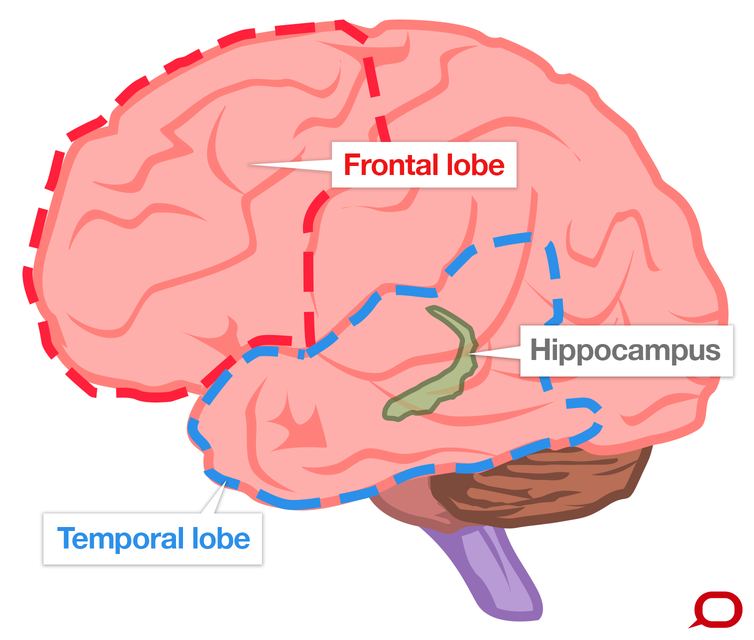
But for a person living with dementia, deterioration in other parts of the braini.e., the frontal lobe, the temporal lobes, the hippocampusgreatly impacts or inhibits processing information.
Unfortunately, our society lacks understanding that these behaviors are influenced by circumstances of day-to-day living. Undesired behaviors can be avoided or reduced when the care provided addresses the persons preferences and wishes. At Hope, we call this person-centered care.
First Some Facts And Figures
Alzheimer’s disease is not a normal part of aging. It is the most common form of dementia, a general term for memory loss and cognitive decline serious enough to interfere with daily life. It’s also, according to the Centers for Disease Control, the 6th top cause of death in the United States. One in three seniors dies with Alzheimer’s or another form of dementia.
Also Check: What Causes Senile Dementia
Six Selected Cases: Tau
To convey how the common neurodegenerative disease-related pathologic markers are distributed in human amygdalae in a small sample, we show digitally rendered low-magnification photomicrographs from 6 human brains . These individuals were followed longitudinally in a community-based cohort from normal clinical status at baseline examination until their eventual autopsy . The autopsies showed pathologic features commonly experienced in this autopsy series . Immunohistochemical stains were used as described previously , along with digital pathologic methods that enable visualization of pathologic markers at low magnification . The consecutive adjacent sections of amygdala were all stained in the same order: , phospho-Tau , -synuclein , and phospho-TDP-43 . The next adjacent section was stained with the modified KlüverBarrera technique to delineate the subfields of the amygdala and peri-amygdaloid regions. Labeling of the amygdalar anatomy was performed based on microscopic review of patient sections stained with a modified KlüverBarrera protocol, incorporating 0.1% Luxol fast blue, Eosin Y, and a 0.5% aqueous solution of Cresyl violet. Anatomic references were used in labeling the KlüverBarrera-stained sections the terminology of Crosby and Humphrey was applied for the amygdalar subnuclei . Note that each immunostain has distinct properties for example, -SN has the highest background, which is true normal staining.
Continued
What Should You Not Say To Someone With Ptsd

10 Things Not to Say to Someone With PTSD What not to say: It wasn’t even life-threatening. … What not to say: People have been through worse. … What not to say: Stop over-reacting. … What not to say: You’re faking it. … What not to say: I’ve been through something similar and I don’t have PTSD, so you don’t have it either.Meer items…15 jan. 2020
Don’t Miss: Vascular Cognitive Impairment Life Expectancy
Key Biological Processes In The Brain
Most neurons have three basic parts: a cell body, multiple dendrites, and an axon.
- The cell body contains the nucleus, which houses the genetic blueprint that directs and regulates the cells activities.
- Dendrites are branch-like structures that extend from the cell body and collect information from other neurons.
- The axon is a cable-like structure at the end of the cell body opposite the dendrites and transmits messages to other neurons.
The function and survival of neurons depend on several key biological processes:
Neurons are a major player in the central nervous system, but other cell types are also key to healthy brain function. In fact, glial cells are by far the most numerous cells in the brain, outnumbering neurons by about 10 to 1. These cells, which come in various formssuch as microglia, astrocytes, and oligodendrocytessurround and support the function and healthy of neurons. For example, microglia protect neurons from physical and chemical damage and are responsible for clearing foreign substances and cellular debris from the brain. To carry out these functions, glial cells often collaborate with blood vessels in the brain. Together, glial and blood vessel cells regulate the delicate balance within the brain to ensure that it functions at its best.
What Are The Side Effects Of Long Term Drinking
Long-Term Health Risks. Over time, excessive alcohol use can lead to the development of chronic diseases and other serious problems including: High blood pressure, heart disease, stroke, liver disease, and digestive problems. Cancer of the breast, mouth, throat, esophagus, voice box, liver, colon, and rectum.
Don’t Miss: What’s The Difference Between Parkinson’s And Alzheimer’s
Vascular Contributions To Alzheimers Disease
People with dementia seldom have only Alzheimers-related changes in their brains. Any number of vascular issuesproblems that affect blood vessels, such as beta-amyloid deposits in brain arteries, atherosclerosis , and mini-strokesmay also be at play.
Vascular problems may lead to reduced blood flow and oxygen to the brain, as well as a breakdown of the blood-brain barrier, which usually protects the brain from harmful agents while allowing in glucose and other necessary factors. In a person with Alzheimers, a faulty blood-brain barrier prevents glucose from reaching the brain and prevents the clearing away of toxic beta-amyloid and tau proteins. This results in inflammation, which adds to vascular problems in the brain. Because it appears that Alzheimers is both a cause and consequence of vascular problems in the brain, researchers are seeking interventions to disrupt this complicated and destructive cycle.
Amygdala Function And Location
By Olivia Guy-Evans, published May 09, 2021
Key Takeaways
- The amygdala in the limbic system plays a key role in how animals assess and respond to environmental threats and challenges byevaluating the emotional importance of sensory information and prompting an appropriate response.
- The main job of the amygdala is toregulate emotions, such as fear and aggression.
- The amygdala is alsoinvolved in tying emotional meaning to our memories.reward processing, and decision-making.
- When it is stimulatedelectrically, animals show aggressive behavior and when it’s removed, they no longer show aggressive behavior.
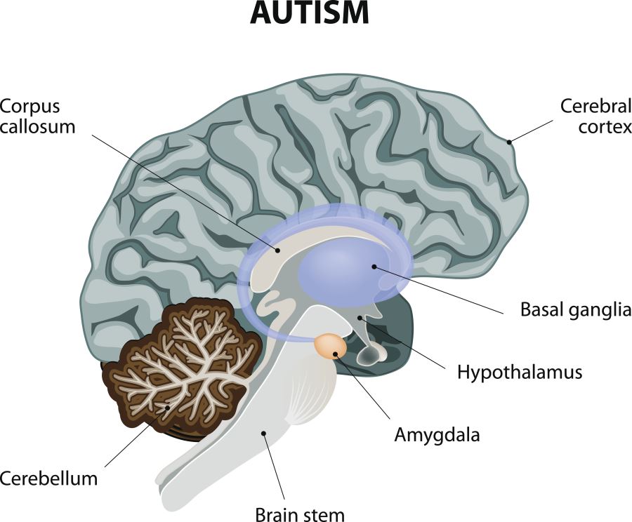
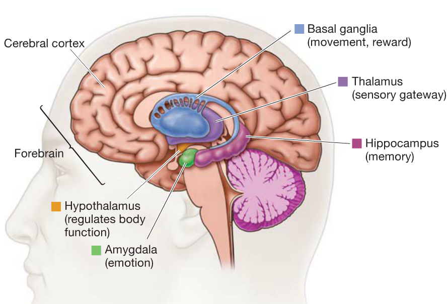
The amygdala is a complex structure of cells nestled in the middle of the brain, adjacent to the hippocampus .
The amygdala is primarily involved in the processing of emotions and memories associated with fear. The amygdala is considered to be a part of the limbic system within the brain and is key to how we process strong emotions like fear or pleasure.
As the amygdala has connections to many other brain structures, this means it can link to areas in order to process âhigherâ cognitive information with systems which control âlowerâ functions .
This allows the amygdala to organize physiological responses based on the cognitive information available.The most well-known example of this is the fight-or-flight response.
There are two amygdalae in each hemisphere of the brain and there are three known functionally distinct parts:
You May Like: Ribbon For Alzheimer’s
The Amygdala & Emotions
The amygdala triggers your emotions faster than your conscious awareness. The unique speed dial circuits of the two almond sized nuclei within your brain are the first to react to emotionally significant events. These organs protect you from harm by interpreting subconscious hints of danger to trigger lightning fast responses. This activity provides more evidence of pattern recognition by the nervous system.
When you imagine that the mind does not compute, but senses patterns, you will be amazed by the incredible competence of these organs. They detect and respond to subliminal signals of danger, or of obstructions to one’s goals. In coordination with the insulae, they also respond with alacrity to negative emotions like grief, guilt, envy, or shame.
The amygdalae react to negative events in many ways, including through the activation of your sympathetic nervous system. The results cause you gut wrenching turmoil. While it takes around 300 milliseconds for you to become aware of a disturbing event, the amygdalae react to it within 20 milliseconds! Sadly, the knee-jerk responses of these organs cause you to overreact to the world around you. Their momentary mischief in the morning can subconsciously trouble you the whole day. An awareness of the mechanism can enable you to effectively still their ill effects and recover your peace of mind.
What Are The Symptoms Of An Amygdala Hijack
The symptoms of an amygdala hijack are caused by the bodys chemical response to stress. When you experience stress, your brain releases two kinds of stress hormones: cortisol and adrenaline. Both of these hormones, which are released by the adrenal glands, prepare your body to fight or to flee.
Together, these stress hormones do a number of things to your body in response to stress. They:
- increase blood flow to muscles, so you have more strength and speed to fight or flee
- expand your airways so you can take in and use more oxygen
- increase blood sugar to provide you immediate energy
- dilate pupils to improve your vision for faster responses
When these hormones are released, you may experience:
- clammy skin
- goosebumps on the surface of your skin
An amygdala hijack may lead to inappropriate or irrational behavior. After an amygdala hijack, you may experience other symptoms like embarrassment and regret.
Read Also: Neurotransmitters Involved In Alzheimer’s Disease
Relation Between Volumes Of Medial Temporal Structures And Memory Performance
Table shows the results of correlation analysis. The right and left amygdala volume and the left subicular volume were significantly correlated with memory test performance. Word recall and logical memory scores correlated with left subicular volume. Verbal paired associates score correlated with right amygdala volume, as did figure memory score with right and left amygdala volumes. Visual reproduction score correlated with the volumes of the right amygdaloid complex and of the left amygdaloid complex. A significant correlation was also found between right hippocampal volume and the visual reproduction score.
Linear univariate regressions of volume of the left subiculum on word recall score and on logical memory score . These relations remained significant after partialling out variance due to age, sex, and education.
How Does The Amygdala Affect Aggression

The amygdala has been shown to be an area that causes aggression. Stimulation of the amygdala results in augmented aggressive behavior, while lesions of this area greatly reduce one’s competitive drive and aggression. Another area, the hypothalamus, is believed to serve a regulatory role in aggression.
Also Check: Did Margaret Thatcher Have Alzheimer’s
What Happens When The Amygdala Is Dysfunctional
The left amygdala has been linked to social anxiety, obsessive and compulsive disorders, and post traumatic stress, as well as more broadly to separation and general anxiety. In a 2003 study, subjects with borderline personality disorder showed significantly greater left amygdala activity than normal control subjects.
What Is The Connection Between The Amygdala And Memory
The amygdala is a structure in the brain usually associated with emotional states. There is, however, a strong connection between the amygdala and memory. Acting in conjunction with other parts of the limbic system, such as the hippocampus, this part of the brain helps regulate and encode emotional memories.
Don’t Miss: Can Dementia Turn Into Alzheimer’s
Atrophy In Basolateral Amygdala In Very Early Ad
Group comparisons revealed decreased amygdala gray matter volumes in AD-MCI and AD-D relative to CON . Slightly lower amygdala volumes in AD-D than in AD-MCI were not significant in the direct contrast. Beyond amygdala, voxel-wise ANOVA revealed a typical pattern of atrophy in AD, including medial and lateral temporal lobes, parieto-occipital regions, insula and cerebellum .
Figure 1. Volumetric group differences across healthy controls , patients with mild cognitive impairment due to Alzheimers disease , and patients with dementia due to Alzheimers disease . shows the main effect of groups with respect to atrophy in gray matter regions Bar represents T-values p< 0.05, FWE corrected. Please cf. to Table S1 in Supplementary Material for details of involved clusters. The y-axis denotes VBM values of amygdala for groups depicted on x-axis. Asterisks indicate significant differences, Bonferroni-corrected p values: CONAD-MCI: p= 0.001 CONAD-D: p< 0.001 AD-MCIAD-D: p= 0.422. Abbreviations: CON, healthy controls AD-MCI, patients with mild cognitive impairment due to Alzheimers disease AD-D, patients with dementia due to Alzheimers disease.
Beyond Emotion: Understanding The Amygdalas Role In Memory
Illustration of the basolateral amygdala , hippocampus , and perirhinal cortex and electrical signals from each region during a recognition test trial. 3-D brain model adapted with permission from AMC Virtual Brain Model. Image courtesy of Cory Inman, Emory University
The amygdalae, a pair of small almond-shaped regions deep in the brain, help regulate emotion and encode memoriesespecially when it comes to more emotional remembrances. Now, new research from Emory University suggests that direct stimulation of the amygdala via deep brain stimulation electrodes can enhance a persons recognition of images seen the day before, leading to the possibility of potential DBS treatment for patients with memory-related disorders.
The amygdala and memory
The amygdala may be best known as the part of the brain that drives the so-called fight or flight response. While it is often associated with the bodys fear and stress responses, it also plays a pivotal role in memory.
One role we are very familiar with, when it comes to the amygdala and memory, is that of emotional salience, says Jon T. Willie, M.D., Ph.D., neurosurgeon and director of the laboratory for behavioral neuromodulation at Emory University in Atlanta. If you have an emotional experience, the amygdala seems to tag that memory in such a way so that it is better remembered.
Stimulating memory, but not emotion
Challenges of DBS as a treatment
Also Check: 7th Stage Of Alzheimer’s
What Happens To The Brain In Alzheimer’s Disease
The healthy human brain contains tens of billions of neuronsspecialized cells that process and transmit information via electrical and chemical signals. They send messages between different parts of the brain, and from the brain to the muscles and organs of the body. Alzheimers disease disrupts this communication among neurons, resulting in loss of function and cell death.
Causes Of Dementia With Lewy Bodies
Lewy bodies are tiny clumps of a protein called alpha-synuclein that can develop inside brain cells.
These clumps damage the way the cells work and communicate with each other, and the brain cells eventually die.
Dementia with Lewy bodies is closely related to Parkinson’s disease and often has some of the same symptoms, including difficulty with movement and a higher risk of falls.
Read more about dementia with Lewy bodies.
Also Check: Familial Dementia
Does Alcohol Affect Memory
Whether its over one night or several years, heavy alcohol use can lead to lapses in memory. This may include difficulty recalling recent events or even an entire night. It can also lead to permanent memory loss, described as dementia. Doctors have identified several ways alcohol affects the brain and memory.
Pathology Of Alzheimers Disease

Macroscopic features
Fig. 1
Gross Anatomy of Alzheimers Brain. Lateral view of an Alzheimers brain can show widening of sulcal spaces and narrowing of gyri compared to a normal brain. This may be more readily observed in coronal sections as indicated by the arrowheads, and this atrophy is often accompanied by enlargement of the frontal and temporal horns of the lateral ventricles as highlighted by the arrows. Additionally, loss of pigmented neurons in the locus coeruleus is commonly observed in the pontine tegmentum as shown with the open circle. None of these features is exclusive to Alzheimers disease
Microscopic features
The definitive diagnosis of AD requires microscopic examination of multiple brain regions employing staining methods that can detect Alzheimer type neuropathologic change , with diagnosis based upon the morphology and density of lesions and their topographic distribution. Several of the brain regions that are vulnerable to Alzheimer type pathologic change are also vulnerable to other disease processes, such as -synucleinopathy and TDP-43 proteinopathy. Mixed pathology is common. Indeed in the Mayo Clinic Brain Bank from 2007 to 2016 , the majority of AD cases had coexisting non-Alzheimer pathologies, and comorbidities increased in frequency with age. Furthermore, when the original clinical diagnoses were examined for cases with pure AD pathology, it is clear that a number of clinical syndromes can masquerade as Alzheimers disease.
Also Check: What Is Senility
Which Part Of The Brain Will Alcohol Affect
The Frontal Lobes: The frontal lobes of our brain are responsible for cognition, thought, memory, and judgment. By inhibiting its effects, alcohol impairs nearly every one of these functions. The hippocampus: The hippocampus forms and stores memory. Alcohols impact on the hippocampus leads to memory loss.