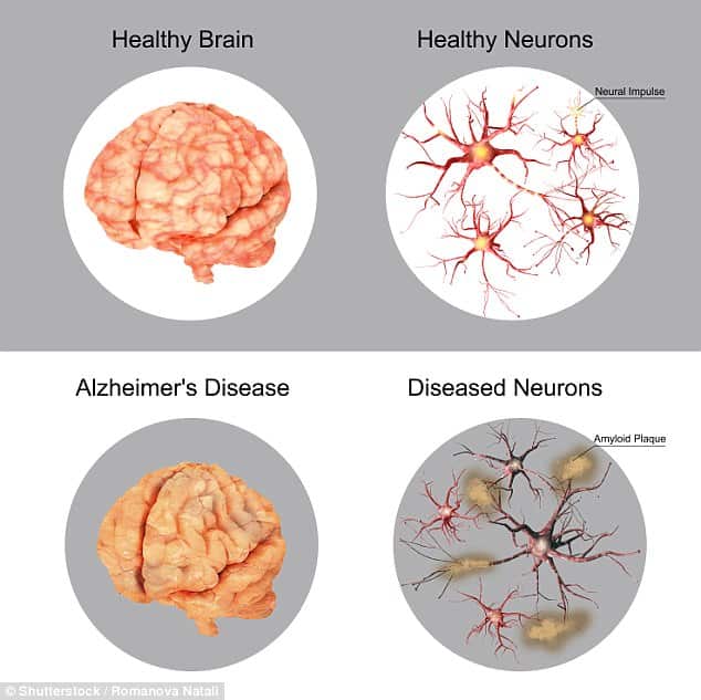
What Parts Of The Brain Are Affected By Dementia
The damaged areas of the brain include the hippocampus, which is an area of the brain that helps new memories form. Damage to the frontal lobe of the brain eventually causes problems with intelligence, judgment, and behaviour. Damage to the temporal lobe affects memory. And damage to the parietal lobe affects language.
Related: Alzheimers Researchers Seethe Over Years Of Missteps After Latest Drug Failure
Since 2012, US health regulators have approved three molecular tracers that bind to amyloid and can be used to visualize it via PET or other neuroimaging, which costs from $3,000 to $7,000 and is not covered by Medicare. Some experts have called for screening everyone older than about 50 for signs of amyloid. But even before this study, research as far back as 1991 showed that many people have amyloid plaques in the brain but have no symptoms of cognitive decline or Alzheimers disease, according to the Alzheimers Association.
Scans can therefore give false positives, said Perry, a longtime skeptic of the idea that amyloid plaques are the chief cause of Alzheimers. The value of the tests as a public health measure is questionable, he said, and the Alzheimers Association does not recommend their routine use to diagnose the disease.
Geulas team studied the brains of eight people older than 90 who were part of the 90+ Study at the University of California, Irvine. Soon before their deaths, the volunteers scored extremely high on memory tests compared to 90-somethings with a normal score. After they died, their brains were scrutinized for telltale signs of Alzheimers.
Three had the characteristic amyloid plaques and tau tangles. But cells in the memory-forming hippocampus and the higher-order-thinking frontal cortex were relatively intact, somehow withstanding the toxic effects of amyloid and tau. The search is on for whats protecting them.
Amyloid Hypothesis Versus Tau Hypothesis
A central but controversial issue in the pathogenesis of AD is the relationship between amyloid deposition and NFT formation. Evidence shows that abnormal amyloid metabolism plays a key pathogenic role. At high concentrations, the fibrillar form of Ab has been shown to be neurotoxic to cultured neurons.
Cultured cortical and hippocampal neurons treated with Ab protein exhibit changes characteristic of apoptosis , including nuclear chromatin condensation, plasma membrane blebbing, and internucleosomal DNA fragmentation. The fibrillar form of Ab has also been shown to alter the phosphorylation state of tau protein.
The identification of several point mutations within the APP gene in some patients with early-onset familial AD and the development of transgenic mice exhibiting cognitive changes and SPs also incriminate Ab in AD. The apolipoprotein E E4 allele, which has been linked with significantly increased risk for developing AD, may promote inability to suppress production of amyloid, increased production of amyloid, or impaired clearance of amyloid with collection outside of the neuron.
Autopsies have shown that patients with 1 or 2 copies of the APOE E4 allele tend to have more amyloid. Additional evidence comes from recent experimental data supporting the role of presenilins in Ab metabolism, as well as findings of abnormal production of Ab protein in presenilin-mutation familial Alzheimer disease.
Recommended Reading: Is Alzheimer’s A Prion Disease
Neurofibrillary Tangles And Senile Plaques
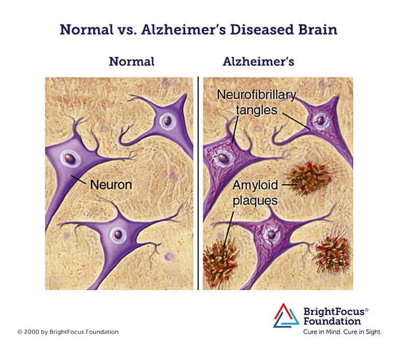
Plaques are dense, mostly insoluble deposits of protein and cellular material outside and around the neurons. Plaques are made of beta-amyloid , a protein fragment snipped from a larger protein called amyloid precursor protein . These fragments clump together and are mixed with other molecules, neurons, and non-nerve cells .
In AD, plaques develop in the hippocampus, a structure deep in the brain that helps to encode memories, and in other areas of the cerebral cortex that are used in thinking and making decisions. Plaques may begin to develop as early as the fifth decade of life. Whether Ab plaques themselves cause AD or whether they are a by-product of the AD process is still unknown. It is known that changes in APP structure can cause a rare, inherited form of AD.
Tangles are insoluble twisted fibers that build up inside the nerve cell. Although many older people develop some plaques and tangles, the brains of people with AD have them to a greater extent, especially in certain regions of the brain that are important in memory. There are likely to be significant age-related differences in the extent to which the presence of plaques and tangles are indicative of the presence of dementia.
SPs also accumulate primarily in association cortices and in the hippocampus. Plaques and tangles have relatively discrete and stereotypical patterns of laminar distribution in the cerebral cortex, which indicate predominant involvement of corticocortical connections.
Estimations Of Amyloid Plaque Density And Volume

The number of A-ir plaques per volume was also estimated by the Optical Fractionator tool in DG, CA3, CA1 and subiculum . A minimum of six sections were selected for each patient, with equal intervals with an ×40 objective on a surface of 22,500 m2 and with a dissector depth of 10 m, rendering a study volume of 225,000 m3 per optical dissector.
To estimate the A-ir plaque volume, the edges of the plaque were delineated with the Nucleator tool with the aid of Stereo Investigator software . This tool provides the volume of each A-ir plaque analyzed, as well as the relative volume occupied by them in each examined hippocampal subfield to provide the percentage of tissue occupied by A-ir plaques.
Also Check: What Is The Difference Between Dementia And Schizophrenia
Sexual Differences In Incidence
Some studies have reported a higher risk of AD in women than in men other studies, however, including the Aging, Demographics, and Memory Study, found no difference in risk between men and women. Almost two thirds of Americans with AD are women. Among AD patients overall, any sexual disparity may simply reflect womens higher life expectancy. Among those who are heterozygous for the APOE E4 allele, however, Payami et al found a twofold increased risk in women.
What Are Amyloid Plaques
Amyloid is a general term for protein fragments that the body produces normally. Beta amyloid is a protein fragment snipped from an amyloid precursor protein . In a healthy brain, these protein fragments are broken down and eliminated. Amyloid plaques are hard, insoluble accumulations of beta amyloid proteins that clump together between the nerve cells in the brains of Alzheimers disease patients.
Recommended Reading: What Kind Of Doctor Tests For Dementia
Estimation Of Neuronal Densities
Densities of labeled neurons were estimated using a stereological method known as optical dissectors with the aid of Stereo Investigator software , using its Optical Fractionator tool. Neuronal densities, expressed as the number of labeled neurons per volume, were estimated in CA3, CA1, and subiculum, using Nissl-stained sections and NeuN-, PHFTau-AT8- and PHFTau-pS396-immunostained sections. Nissl-stained and NeuN-immunostained sections were used to identify the boundaries within the hippocampus.
After randomly selecting a starting point, six sections were chosen at equally spaced intervals. Optical dissectors were made with an oil immersion ×100 objective for both the NeuN-immunostained and Nissl-stained sections, on an average surface of 2,050 m2. The depth of the optical dissectors was 10 m, rendering a study volume of 20,500 m3 per optical dissector. An ×40 objective was used for the PHFTau-immunostained sections, on a surface of 14,450 m2. The depth of the optical dissectors in this case was also 10 m, rendering a study volume of 144,500 m3. Stereological parameters for each sample and neuronal marker were chosen. Since most neurons are located in the pyramidal cell layer, neuronal densities were estimated in this layer in the CA subfields and subiculum. In Nissl-stained sections, a neuron was only counted if the nucleolus was clearly identified in the optical plane along the vertical z-axis .
Granulovacuolar Degeneration And Neuropil Threads
Granulovacuolar degeneration occurs almost exclusively in the hippocampus. Neuropil threads are an array of dystrophic neurites diffusely distributed in the cortical neuropil, more or less independently of plaques and tangles. This lesion suggests neuropil alterations beyond those merely due to NFTs and SPs and indicates an even more widespread insult to the cortical circuitry than that visualized by studying only plaques and tangles.
Also Check: When Was Dementia First Discovered
How A Good Nights Sleep Could Reduce Plaques And Tangles
Many caregivers report that Alzheimers causes disrupted sleep and sundowning. Sundowning is a common Alzheimers symptom that leads to increased confusion in the late afternoon or early evening. But researchers have also discovered that frequent nights spent tossing and turning may also cause Alzheimers.
In a recent study, scientists found that those with sleep apnea or who snore loudly were more likely to have increased amounts of tau in their brains. Other studies have found that participants with fragmented sleep patternstaking short cat-naps throughout the dayhad more beta-amyloid plaques than participants who got a consistent eight hours of sleep each night. A 2017 analysis found that poor sleepers appeared to have about a 68 percent higher risk of cognitive issues including Alzheimers than those who were well-rested. In other words, getting some shut-eye will not only give your brain a boost, but could also prevent your brain from developing additional plaques and tangles.
Plaques Tangles And Dementia
Although the combination of A plaques and NFTs was soon recognized as a histopathologic hallmark of DAT, the pathogenesis of the development of the clinical syndrome remained unclear for many years. Even Alzheimer acknowledged that these abnormal structures likely were associated with neuronal dysfunction however, it was uncertain at the time whether the histopathologic findings were cause or effect.,,, To this day, the relationship between plaques and tangles is also incompletely understood. In Alzheimer’s second patient, no NFTs were noted, leading some investigators to consider that A pathology may precede and even promote pathologies., To date, however, no proved mechanisms directly link the formation of NFTs to A pathology.,
Fig 2.
Diagrammatic representation of APP processing. APP is a transmembrane protein with both extra- and intracellular components. APP is processed by 2 competing pathways. The -secretase pathway generates sAPP and C83 protein by cleavage of the -secretase enzyme . In the -secretase pathway, the enzyme -secretase cleaves APP into an sAPP fragment and a C99 fragment. The C99 fragment is further cleaved by -secretase enzyme into an amyloid fragment and an AICD fragment. The A fragments polymerize. The oligomers and polymers exhibit neurotoxicity. As polymerization proceeds to more complex forms, senile plaques are developed. The C83 fragment is also further processed. However, the function of its products is not fully understood.
Don’t Miss: What Is Dementia Teepa Snow
Do Plaques Cause Alzheimer’s
So far, the prevailing hypothesis among experts has been that the excessive accumulation of a potentially toxic protein beta-amyloid in the brain causes Alzheimer’s. Researchers have argued that beta-amyloid plaques disrupt the communication between brain cells, potentially leading to cognitive function problems.
What Causes Neurofibrillary Tangles To Form

Tangles form when tau is misfolded in a very specific way. In Alzheimers disease, the tau forms a C-shape in the core of the tangle with a loose end sticking out randomly. In Picks disease, the core forms a J-shape instead. Once a tangle has been started, more tau proteins are recruited to make it longer. There is likely a molecule responsible for shaping tau into these forms, but it has not yet been identified.
Don’t Miss: Can A Person With Dementia Have Hallucinations
Plaques Tangles And Imaging
As mechanisms for the development of AD become better understood, clinical emphasis will increasingly focus on early diagnosis and evaluation of proposed treatments. Attention has recently concentrated on the condition of mild cognitive impairment and its relationship to overt dementia. Limitations of standard psychometric tools have prompted some investigators to consider imaging as a proxy for determining clinical outcomes. Indeed, the National Institute on Aging and the National Institute of Biomedical Imaging and Bioengineering in partnership with several pharmaceutical firms and national foundations have formed the Alzheimer Disease Neuroimaging Initiative to explore new imaging techniques and optimize methods for image acquisition in longitudinal studies. In vivo imaging of cerebral amyloid by using molecular probes and PET, volumetric analysis of select portions of the brain with high-resolution MR imaging, and diffusion tensor imaging are just a few of the exciting applications of neuroimaging that will develop in the years to come.
Cholinergic Neurotransmission And Alzheimer Disease
The cholinergic system is involved in memory function, and cholinergic deficiency has been implicated in the cognitive decline and behavioral changes of AD. Activity of the synthetic enzyme choline acetyltransferase and the catabolic enzyme acetylcholinesterase are significantly reduced in the cerebral cortex, hippocampus, and amygdala in patients with AD.
The nucleus basalis of Meynert and diagonal band of Broca provide the main cholinergic input to the hippocampus, amygdala, and neocortex, which are lost in patients with AD. Loss of cortical CAT and decline in acetylcholine synthesis in biopsy specimens have been found to correlate with cognitive impairment and reaction-time performance. Because cholinergic dysfunction may contribute to the symptoms of patients with AD, enhancing cholinergic neurotransmission constitutes a rational basis for symptomatic treatment.
Recommended Reading: How Do You Avoid Dementia
What Causes Beta Amyloid Plaques
Beta amyloid molecules are initially found in very small strands that can dissolve in the fluid between cells, which will be washed out of the brain. However, the enzyme that cuts APP into beta amyloid is not very precise and can also result in slightly larger strands that do not dissolve. The longer strands are very sticky at the level of individual molecules and start the process of clumping into the deposits referred to as plaques.
The Connection Between Exercise And Brain Plaque Reduction
Scientists reported that when we exercise, our brains produce a protein known as Irisin, which benefits our thinking abilities and may even lead to neural growth in the hippocampus. In another recent study, scientists found that aerobic exercise could make your brain act up to 20 years younger. The benefits of exercise dont end there: Researchers think that in addition to sleep, exercising could also decrease Alzheimers-related plaques and tangles in the brain.
Last year, scientists pushed mice genetically engineered to have Alzheimers to exercise for up to three hours at a time by giving them running wheels. They gave the other Alzheimers mice gene therapy and drugs meant to help them form new nerve cells. The researchers learned that the mice who exercised did better on cognitive tests than the other group and had smaller levels of beta-amyloid plaques in their brains. Neuroscientist Wendy Suzuki said that exercise can alsore-sculpt your brain and lower dementia risk by up to 90 percent.
Also Check: How To Talk To A Patient With Dementia
Age Distribution For Alzheimer Disease
The prevalence of AD increases with age. AD is most prevalent in individuals older than 60 years. Some forms of familial early-onset AD can appear as early as the third decade, but familial cases constitute less than 10% of AD overall.
More than 90% of cases of AD are sporadic and occur in individuals older than 60 years. Of interest, however, results of some studies of nonagenarians and centenarians suggest that the risk may decrease in individuals older than 90 years. If so, age is not an unqualified risk factor for the disease, but further study of this matter is needed.
Savva et al found that in the elderly population, the association between dementia and the pathological features of AD is stronger in persons 75 years of age than in persons 95 years of age. These results were achieved by assessing 456 brains donated to the population-based Medical Research Council Cognitive Function and Ageing Study from persons 69-103 years of age at death.
Studies have demonstrated that the relationship between cerebral atrophy and dementia persist into the oldest ages but that the strength of association between pathological features of AD and clinical dementia diminishes. It is important to take age into account when assessing the likely effect of interventions against dementia.
Oxidative Stress And Damage
Oxidative damage occurs in AD. Studies have demonstrated that an increase in oxidative damage selectively occurs within the brain regions involved in regulating cognitive performance.
Oxidative damage potentially serves as an early event that then initiates the development of cognitive disturbances and pathological features observed in AD. A decline in protein synthesis capabilities occurs in the same brain regions that exhibit increased levels of oxidative damage in patients with mild cognitive impairment and AD. Protein synthesis may be one of the earliest cellular processes disrupted by oxidative damage in AD.
Oxidative stress is believed to be a critical factor in normal aging and in neurodegenerative diseases such as Parkinson disease, amyotrophic lateral sclerosis, and AD. Formation of free carbonyls and thiobarbituric acid-reactive products, an index of oxidative damage, are significantly increased in AD brain tissue compared with age-matched controls. Plaques and tangles display immunoreactivity to antioxidant enzymes.
Multiple mechanisms exist by which cellular alterations may be induced by oxidative stress, including production of reactive oxygen species in the cell membrane . This in turn impairs the various membrane proteins involved in ion homeostasis such as N -methyl-D-aspartate receptor channels or ion-motive adenosine triphosphatases.
You May Like: What Do Alzheimer’s Patients Think About
How Does Alzheimers Disease Affect The Brain
The brain typically shrinks to some degree in healthy aging but, surprisingly, does not lose neurons in large numbers. In Alzheimers disease, however, damage is widespread, as many neurons stop functioning, lose connections with other neurons, and die. Alzheimers disrupts processes vital to neurons and their networks, including communication, metabolism, and repair.
At first, Alzheimers disease typically destroys neurons and their connections in parts of the brain involved in memory, including the entorhinal cortex and hippocampus. It later affects areas in the cerebral cortex responsible for language, reasoning, and social behavior. Eventually, many other areas of the brain are damaged. Over time, a person with Alzheimers gradually loses his or her ability to live and function independently. Ultimately, the disease is fatal.